
Magnetic immuno-chromatographic test paper strip for quantitatively detecting carcinoembryonic antigen in blood and preparation method thereof

A technology of magnetic immunochromatography and carcinoembryonic antigen, which is applied in the field of medical testing, can solve problems such as expensive instruments and equipment, hazards to operators, and difficulty in marketing, and achieve the effect of simplifying the preparation process and easy operation

Inactive Publication Date: 2010-06-30
CHEMCLIN DIAGNOSTICS CO LTD
View PDF5 Cites 11 Cited by
  • Summary
  • Abstract
  • Description
  • Claims
  • Application Information

AI Technical Summary

Problems solved by technology

[0003] The current diagnostic techniques for CEA in blood mainly include: radioimmunoassay (RIA), enzyme-linked immunoassay (ELISA), chemiluminescence (CLIA), time-resolved fluorescence, colloidal gold immunochromatography and other methods, but these methods have their own The characteristics and deficiencies of radioimmunoassay: the results of radioimmunoassay are relatively accurate and the linear range is wide, but the operation is cumbersome and time-consuming, and the radioactive markers will cause harm to the operator and cause environmental pollution. At present, it has been gradually replaced by other methods; The principle of ELISA is similar to that of chemiluminescence and time-resolved fluorescence, but there is a difference in sensitivity due to the difference in the final detection system. At present, automation, large-scale, and quantitative detection have been realized, but automated detection is currently monopolized by large foreign manufacturers. The instruments and equipment are expensive, and they are not suitable for single-person and small batch testing, which greatly limits their application in primary hospitals and clinics
In recent years, colloidal gold immunochromatography has appeared to detect CEA, which is fast, convenient, and single-serve operation, but the disadvantage is that it can only achieve semi-quantitative, can only indicate a rough range, and cannot achieve accurate quantification, and abnormal samples need to use other methods Double-check testing will virtually increase the cost of medical treatment for the testees, and it is difficult to promote in the market, and it is difficult to use it on a large scale

Method used


Image

  • Magnetic immuno-chromatographic test paper strip for quantitatively detecting carcinoembryonic antigen in blood and preparation method thereof
  • Magnetic immuno-chromatographic test paper strip for quantitatively detecting carcinoembryonic antigen in blood and preparation method thereof

Examples


Embodiment 1

[0033] Magnetic immunochromatographic test strip for quantitatively detecting carcinoembryonic antigen in blood and preparation method of test box

[0034] The preparation method of the test strip and the test carton of this embodiment includes the following steps:

[0035] A. Antibody preparation: select a commercial CEA paired antibody (Hytest, cat: 4CA30), and dialyze it overnight at 4°C with 20mM, pH7.2 (applicable to pH7.0-7.6) PBS.

[0036] B. Preparation of coating film:

[0037] Preparation of coating buffer: 0.02M pH 7.2 phosphate buffer (PB) is the coating buffer, 0.22μm microporous membrane filter sterilizes and store at 4°C for later use. The validity period is two weeks.

[0038] The preparation of the blocking solution: 0.02M phosphate buffered saline (PBS) containing 0.5% BSA, pH 7.2 (pH 7.0-7.6 are applicable), 0.22μm microporous membrane filter sterilized and stored at 4°C for later use , Valid for one week.

[0039] Preparation of coating film: use coating buffer (0.02...

Embodiment 2

[0068] Except for the steps in the preparation of streptavidinized magnetic particles: streptavidin makes the ratio of streptavidin to magnetic particles 1:3, the other steps are the same as in Example 1.

Embodiment 3

[0070] Except for the steps in the preparation of streptavidinized magnetic particles: streptavidin makes the ratio of streptavidin to magnetic particles 1:8, the other steps are the same as in Example 1.


PUM


Abstract

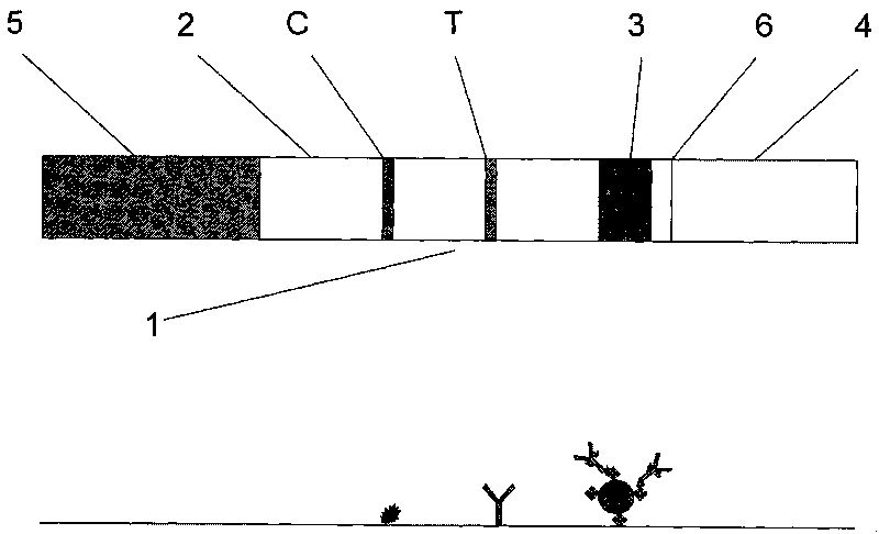
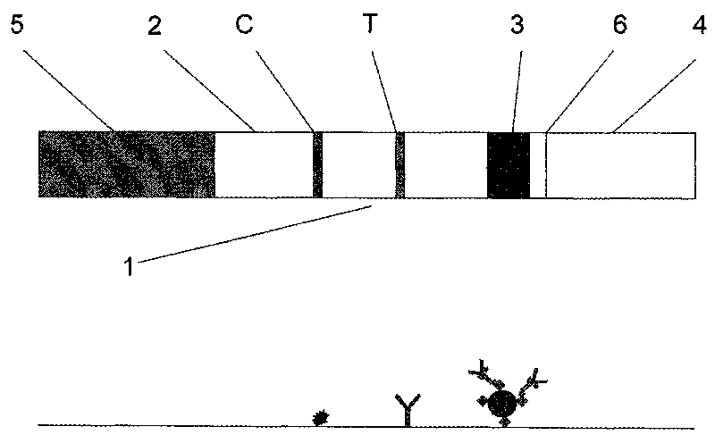
The invention relates to a magnetic immuno-chromatographic test paper strip for quantitatively detecting carcinoembryonic antigen in blood and a preparation method thereof. A coating film, a magnet particle pad combined with CEA antibodies, a sample pad and a water absorbing pad are sequentially and alternately stuck on a base plate in a staggering way at intervals of 2mm, and then the upper layer is covered by a transparent plastic sealing film to form a test paper strip. A CEA antibody detection line and a quality control line are pre-coated on the coating film. The invention introduces the magnetic immuno-chromatographic technology and the biotin-aviden system into the quantitative detection of CEA in blood, and has the advantages that the detection sensitivity is greatly improved, the accurate quantitative results can be given, the labor strength of the operators is reduced, and the clinicians can rapidly and accurately diagnose the illness of patients.

Description

Technical field [0001] The invention relates to a magnetic immunochromatographic test strip for quantitatively detecting carcinoembryonic antigen in blood in the field of medical testing and a preparation method. Background technique [0002] CEA is a globulin that was first discovered in the serum of colon cancer patients by Phil Gold and Samuel O. Freedman in 1965. It was subsequently discovered in colon, stomach, pancreatic, lung, and breast cancer patients. It is produced during embryonic development and stopped after birth, so it is called carcinoembryonic antigen. The content of CEA in normal adults is very low and will only increase under certain pathological conditions (tumor or inflammation). The main clinical significance of carcinoembryonic antigen as a tumor marker for diagnosis is as follows: (1) Patients with primary colon cancer The increase in CEA accounts for 45-80%. (2) In addition to primary colon cancer, adenopancreatic cancer, cholangiocarcinoma, and gastri...

Claims


Application Information

Patent Timeline
IPC IPC(8): G01N33/574G01N33/531G01N33/532G01N33/577G01N33/558
Inventor 姚洪涛应希堂李强胡国茂郑金来张坤
Owner CHEMCLIN DIAGNOSTICS CO LTD
Learn More