
Intravascular photoacoustic and ultrasonic double-mode imaging endoscope device and imaging method thereof

A dual-mode imaging and endoscope technology, applied in catheters, medical science, sensors, etc., can solve the problem of shallow penetration depth and achieve the effect of convenient use and small size

Active Publication Date: 2010-12-15
SOUTH CHINA NORMAL UNIVERSITY
View PDF3 Cites 72 Cited by
  • Summary
  • Abstract
  • Description
  • Claims
  • Application Information

AI Technical Summary

Problems solved by technology

The basic principles of the above three types of endoscopic technologies are to use optical scattering or reflection signals to obtain detection images. Due to the strong influence of medium scattering, there is a shortcoming of shallow penetration depth (~ mm level)

Method used


Image

  • Intravascular photoacoustic and ultrasonic double-mode imaging endoscope device and imaging method thereof
  • Intravascular photoacoustic and ultrasonic double-mode imaging endoscope device and imaging method thereof
  • Intravascular photoacoustic and ultrasonic double-mode imaging endoscope device and imaging method thereof

Examples


Embodiment 1

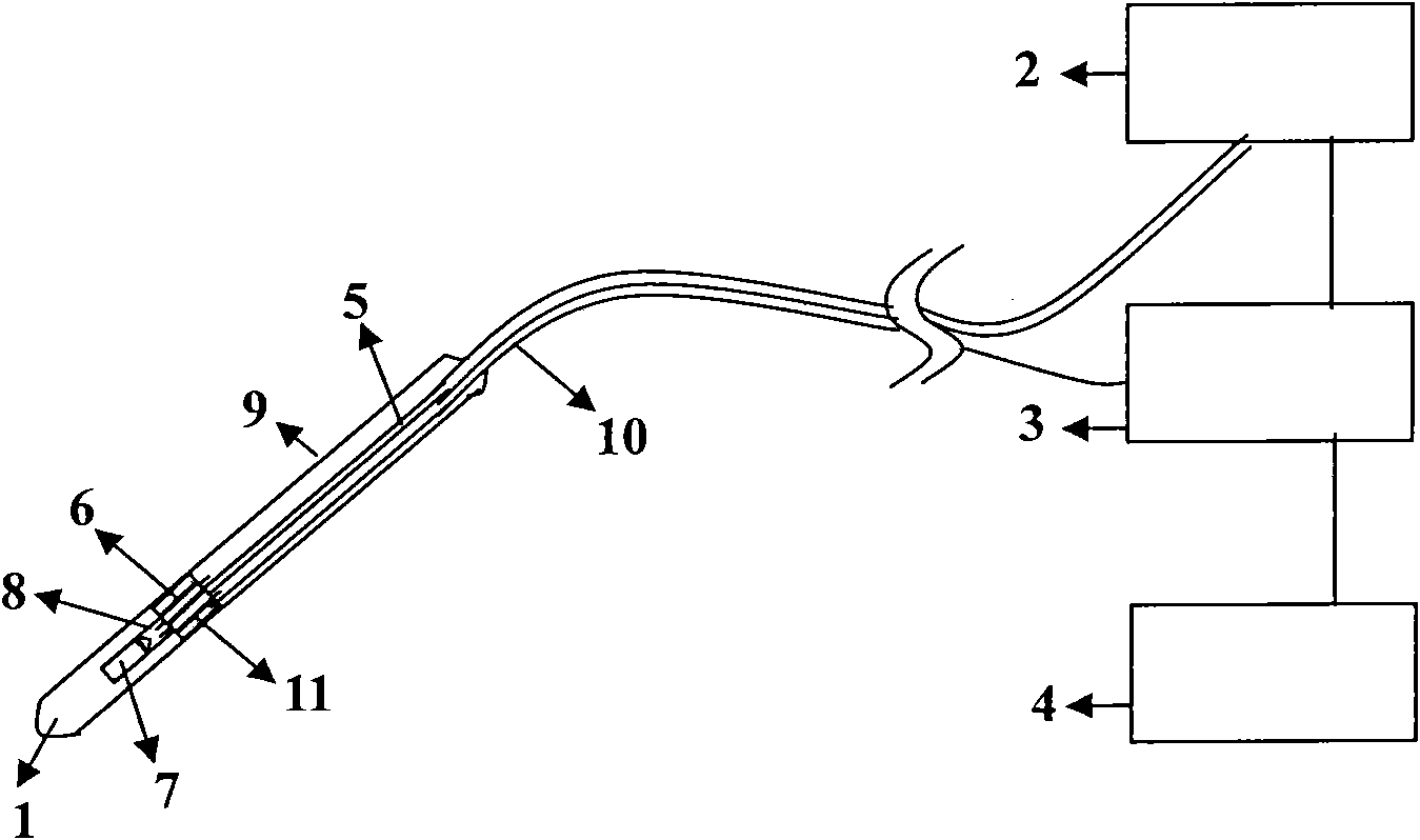
[0034] Example 1: A dual-mode endoscopic imaging device for intravascular photoacoustic ultrasound (structural schematic diagram as shown in figure 1 shown)

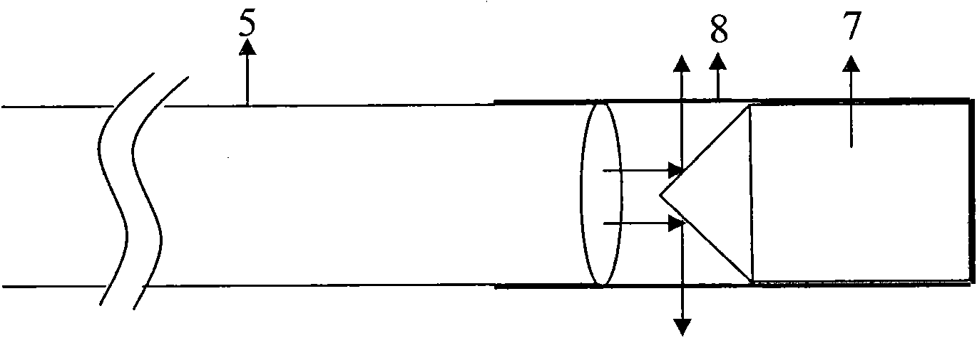
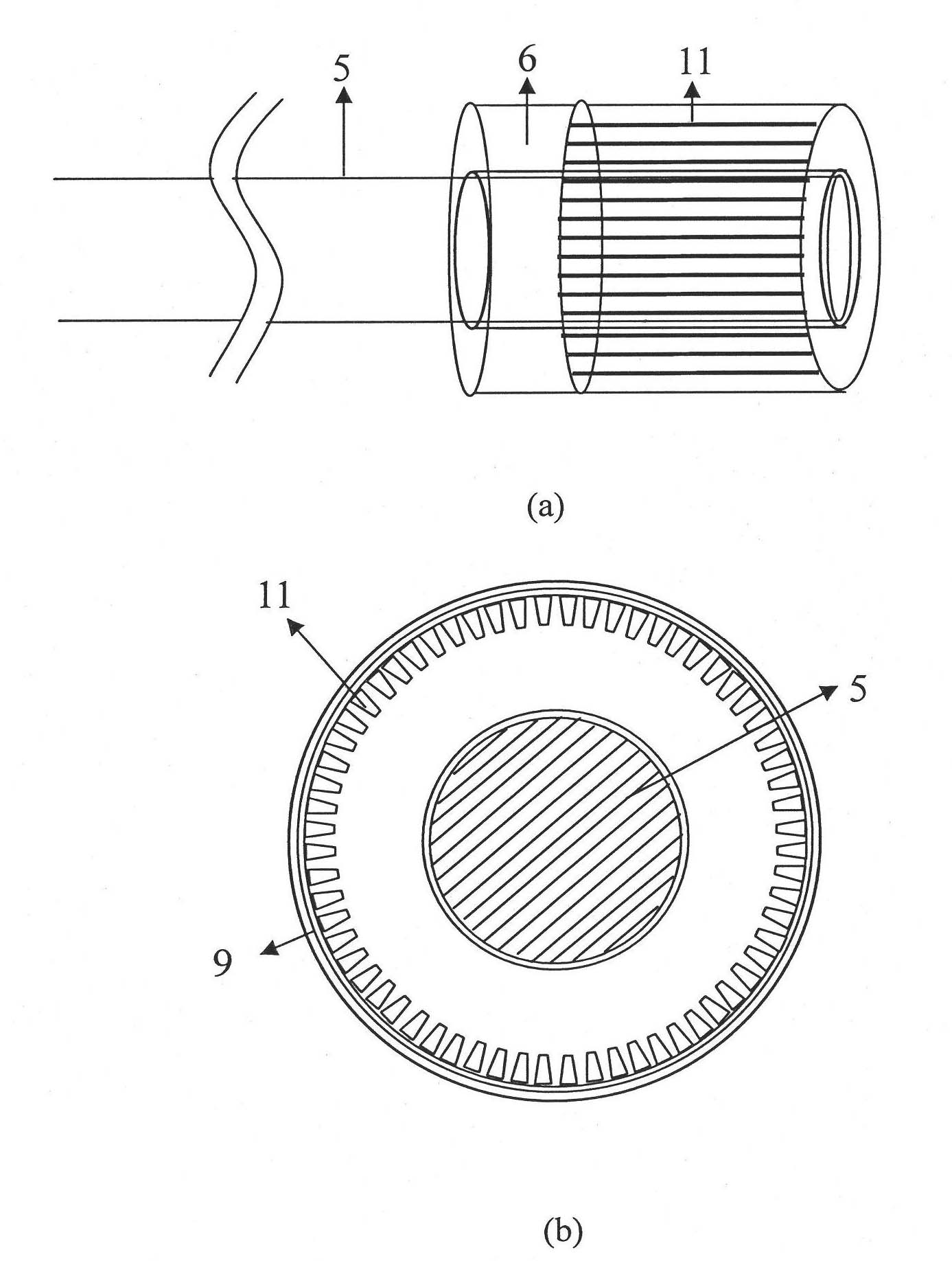
[0035]The device includes a miniature photoacoustic ultrasound dual-mode imaging endoscopic probe 1, a photoacoustic excitation light source generator 2, an ultrasonic signal excitation and acquisition component 3, and a computer 4 with acquisition control software and image reconstruction software; the miniature photoacoustic ultrasound A dual-mode imaging endoscopic probe 1, a photoacoustic excitation light source generator 2, an ultrasonic signal excitation and acquisition component 3, and a computer 4 with acquisition control software and image reconstruction software are electrically connected in sequence; the miniature photoacoustic ultrasonic dual-mode imaging The peeping probe 1 is composed of an incident optical fiber 5, a hollow circular ring array ultrasonic probe 6, a coating tapered high reflection mirror 7,...

Embodiment 3

[0044] Application of the device described in Example 1 to realize photoacoustic and ultrasonic endoscopic imaging of the cross-section of the vascular lumen

[0045] (1) Insert a miniature photoacoustic ultrasound dual-mode imaging endoscopic probe (1.3 mm in diameter) into the lumen of the isolated rabbit abdominal aorta;

[0046] (2) Photoacoustic excitation, photoacoustic collection, and ultrasonic excitation and collection: a fiber laser (Brilliant B, Bigsky) pumped by Nd:YAG is used, the output laser wavelength is 1064nm, the pulse width is 8ns, and the repetition frequency is 20HZ; The pulsed laser (with a wavelength range of 1000nm) generated by the exciter is coupled through the incident optical fiber, and is uniformly incident on the wall of the blood vessel laterally through the coated conical high-reflection mirror to excite the blood vessel wall to generate photoacoustic signals; the hollow ring array ultrasonic detection After receiving the photoacoustic signal, ...


PUM

PropertyMeasurementUnit
Diameteraaaaaaaaaa
Wavelengthaaaaaaaaaa
Pulse widthaaaaaaaaaa

Abstract

The invention discloses an intravascular photoacoustic and ultrasonic double-mode imaging endoscope device and an imaging method thereof. The device comprises a photoacoustic excitation light source generator, a micro photoacoustic and ultrasonic double-mode endoscope probe, an ultrasonic signal exciting and acquiring subassembly and a computer, wherein the micro photoacoustic and ultrasonic double-mode endoscope probe consists of an incident optical fiber, a hollow annular array ultrasonic detector, a coated conical surface high reflector, a tube shell and a PVC tube; the incident optical fiber is connected with the photoacoustic excitation light source generator and passes through the hollow annular array ultrasonic detector; the hollow annular array ultrasonic detector is connected with the ultrasonic signal exciting and acquiring subassembly through a coaxial data transmission line; the coated conical surface high reflector is fixed in the tube shell; and the tube shell is fixedly connected with the outlet of the optical fiber so that the output laser is reflected circularly on the coated conical surface outwards and matched with the hollow annular array ultrasonic detector. The device and the method can realize imaging of a photoacoustic and ultrasonic double-mode endoscope and can be applied to the photoacoustic and ultrasonic double-mode imaging in vessels or micro cavities.

Description

technical field [0001] The invention belongs to the non-destructive testing and measuring technology, and in particular relates to an endoscopic imaging device and an imaging method thereof which use photoacoustic ultrasonic dual-mode imaging technology for intravascular structure imaging. Background technique [0002] Endoscope is a multi-disciplinary and general-purpose tool. Its function is to probe into the depths of curved pipes, to observe parts that cannot be seen directly, to observe the internal space structure and state in a sealed cavity, and to realize long-distance Observation and operation. The currently commonly used optical endoscopes are mainly NDT instruments integrating light, machinery and electricity, which are divided into three series of products: the first category, rigid endoscope series; the second category, fiber endoscope series; the third category Three types, electronic video endoscope series. The working principle of the rigid endoscope serie...

Claims


Application Information

Patent Timeline
IPC IPC(8): A61B1/00A61B5/00A61B8/12
Inventor 邢达袁毅杨思华
Owner SOUTH CHINA NORMAL UNIVERSITY
Features
  • Generate Ideas
  • Intellectual Property
  • Life Sciences
  • Materials
  • Tech Scout
  • Unparalleled Data Quality
  • Higher Quality Content
  • 60% Fewer Hallucinations
Learn More