Combined detection method and diagnostic kit of fusion genes related to lymphoma

A diagnostic kit and fusion gene technology, which are applied in the determination/examination of microorganisms, biochemical equipment and methods, fluorescence/phosphorescence, etc., can solve the complex operation of fluorescence in situ hybridization technology, can not really meet the clinical diagnosis and detection, and have insufficient sensitivity. Good and other problems, to achieve the effect of high specificity, rapid detection, and high sensitivity

Active Publication Date: 2011-01-26
MEDI GENETECH
View PDF3 Cites 12 Cited by
  • Summary
  • Abstract
  • Description
  • Claims
  • Application Information

AI Technical Summary

Problems solved by technology

[0005] Among the currently widely used molecular biology methods for detecting lymphoma fusion genes at home and abroad, fluorescence in situ hybridization (FISH) can only perform qualitative detection, and the operation is complicated; fluorescent quantitative PCR has lim

Method used


Image

  • Combined detection method and diagnostic kit of fusion genes related to lymphoma
  • Combined detection method and diagnostic kit of fusion genes related to lymphoma
  • Combined detection method and diagnostic kit of fusion genes related to lymphoma

Examples


Embodiment 1

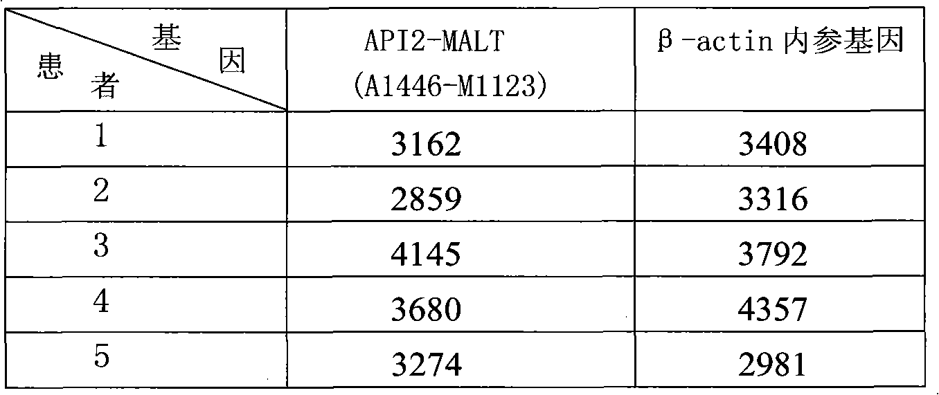
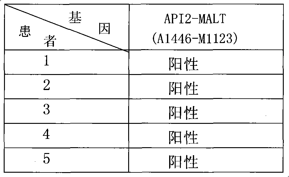
[0067] Example 1: A liquid-phase chip detection method for a lymphoma-related fusion gene

[0068] The specific detection method includes the following steps:

[0069] 1. Preparation of microsphere mixture for detection of API2-MALT1(A1446-M1123) fusion gene

[0070] 1. Synthesize oligonucleotide probes according to the following sequence:

[0071] API2-MALT1 (A1446-M1123): 5'-AminolinkerC12 CCAAGATTATTTAATTCATTTG-3', as shown in SEQ ID NO.2;

[0072] β-actin gene: 5'-AminolinkerC12 TCATTGTAGAAGGTGTGGTG-3', as shown in SEQ ID NO.15;

[0073] 2. Coupling the oligonucleotide probe containing amino modification with two kinds of carboxyl microspheres numbered 25 and 64 respectively

[0074] 2.1 Take out a small portion of fresh dry powdered EDC stored at -20°C and equilibrate to room temperature;

[0075] 2.2 with dH 2 O dissolve the oligonucleotide probes of API2-MALT1 (A1446-M1123) and β-actin respectively at a concentration of 1 mM (1 nmol / μl);

[0076] 2.3 Vortex the st...

Embodiment 2

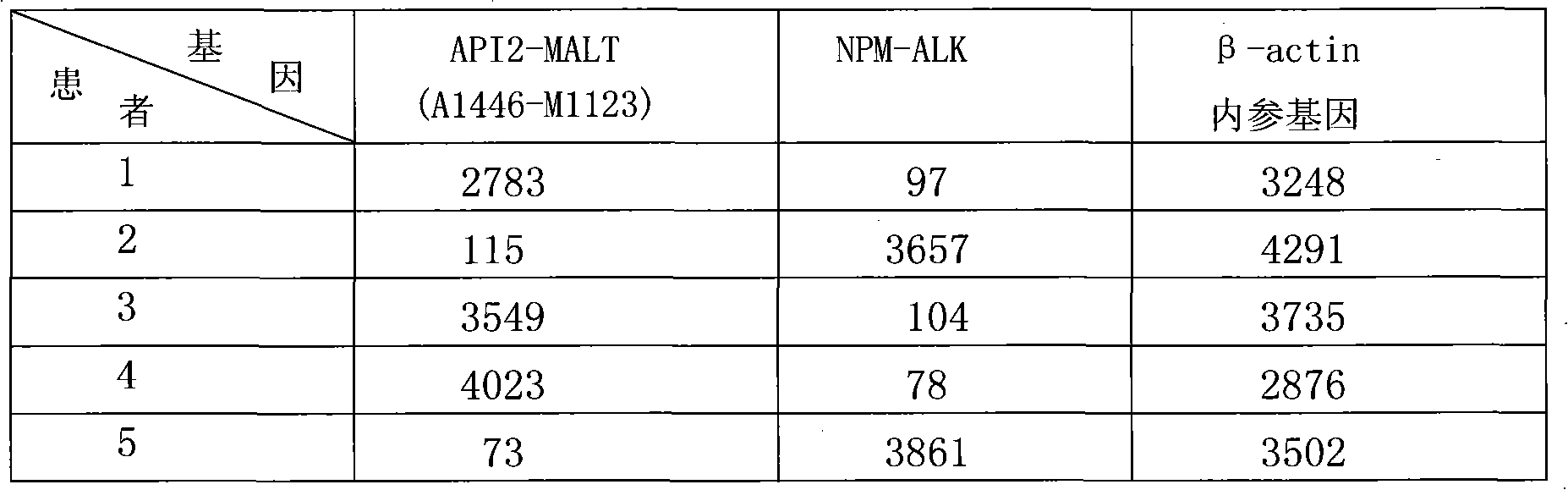
[0169] Example 2: Liquid-phase chip combined detection method for two lymphoma-related fusion genes

[0170] The specific detection method includes the following steps:

[0171] 1. Preparation of microsphere mixture for detection of API2-MALT1 (A1446-M1123), NPM-ALK fusion gene

[0172] 1. Synthesize oligonucleotide probes according to the following sequence:

[0173] API2-MALT1 (A1446-M1123): 5'-AminolinkerC12 CCAAGATTATTTAATTCATTTG-3', as shown in SEQ ID NO.2;

[0174] NPM-ALK: 5'-AminolinkerC12 CCTATAGTTGTTTTAAATGC-3', as shown in SEQ ID NO.4;

[0175] β-actin gene: 5'-AminolinkerC12 TCATTGTAGAAGGTGTGGTG-3', as shown in SEQ ID NO.15;

[0176] 2. Coupling the oligonucleotide probes containing amino modifications to three kinds of carboxyl microspheres numbered 25, 50, and 64 respectively

[0177] 2.1 Take out a small portion of fresh dry powdered EDC stored at -20°C and equilibrate to room temperature;

[0178] 2.2 with dH 2 O dissolve the oligonucleotide probes of API...

Embodiment 3

[0273] Example 3: Liquid-phase chip combined detection method for 3 lymphoma-related fusion genes

[0274] The specific detection method includes the following steps:

[0275] 1. Preparation of microsphere mixture for detection of API2-MALT1 (A1446-M814), API2-MALT1 (A1446-M1123), and NPM-ALK fusion genes

[0276] 1. Synthesize oligonucleotide probes according to the following sequence:

[0277] API2-MALT1 (A1446-M814): 5'-AminolinkerC12 GCTTTGATTCTTTTTTCTCAG-3', as shown in SEQ ID NO.1;

[0278] API2-MALT1 (A1446-M1123): 5'-AminolinkerC12 CCAAGATTATTTAATTCATTTG-3', as shown in SEQ ID NO.2;

[0279] NPM-ALK: 5'-AminolinkerC12 CCTATAGTTGTTTTAAATGC-3', as shown in SEQ ID NO.4;

[0280] β-actin gene: 5'-AminolinkerC12 TCATTGTAGAAGGTGTGGTG-3', as shown in SEQ ID NO.15;

[0281] 2. Coupling the oligonucleotide probes containing amino modifications to four kinds of carboxyl microspheres numbered 11, 25, 50, and 64 respectively

[0282] 2.1 Take out a small portion of fresh dry ...


PUM


Abstract

The invention discloses a combined detection method and a diagnostic kit of fusion genes related to lymphoma. By designing primers and probes for fusion genes API2-MALT1(A1446-M814), API2-MALT1(A1446-M1123), API2-MALT1(A1446-M1150) and NPM-ALK related to lymphoma, a probe and microsphere mixture formed by covalent combination of the probes and the microspheres is hybridized with a reverse transcription-polymerase chain reaction (RT-PCR) amplification product, and after streptavidin phycoerythrin is added, fluorescence signals of different microspheres can be detected, thereby determining whether the sample to be detected contains the fusion genes related to lymphoma or not and determining the expression conditions of the fusion genes. The method and the kit of the invention have the advantages of high sensitivity, high flux, quick and accurate detection and the like, and can be used for qualitatively and quantitatively detecting various lymphoma fusion genes simultaneously.

Description

technical field [0001] The invention relates to the technical field of in vitro diagnosis and detection, in particular to a combined liquid phase chip detection method and a diagnostic kit for multiple lymphoma-related fusion genes. Background technique [0002] Lymphoma is a group of malignant tumors originating from lymph nodes or other lymphoid tissues. It can be divided into two categories: Hodgkin's disease (HD) and non-Hodgkin's lymphoma (NHL). one. Histologically, tumorous proliferation of lymphocytes and (or) histiocytes can be seen, and the main clinical manifestation is painless and progressive lymphadenopathy. [0003] Mucosa-associated lymphoid tissue (MALT) lymphoma is a malignant lymphoma that originates outside lymph nodes, and its incidence accounts for about 40% of all lymphomas, among which MALT lymphoma of the gastrointestinal tract is the most common. API2-MALT1 is a fusion gene produced by chromosomal translocation in MALT lymphoma, which can be found ...

Claims


Application Information

Patent Timeline
IPC IPC(8): C12Q1/68G01N21/64
CPCC12Q2600/16C12Q1/6886C12Q2600/158
Inventor 邵棠袁福美
Owner MEDI GENETECH
Why Eureka
Try Eureka