Electrochemical immunodetection method
An immunodetection method and electrochemical technology are applied in the field of electrochemical immunodetection of thionine-graphene nanocomposite, which can solve the problem that it is difficult to detect trace abnormal prothrombin, affects the accuracy of primary liver cancer, and has low sensitivity. question
- Summary
- Abstract
- Description
- Claims
- Application Information
AI Technical Summary
Problems solved by technology
Method used
Image
Examples
Embodiment 1
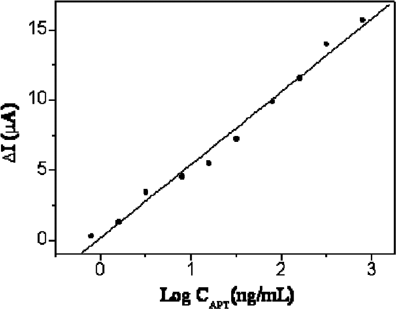
[0046] The preparation of the nanocomposite of embodiment 1 nano-platinum chitosan double wrapping thionine-graphene
[0047] Add 5 mg of thionine to 4.5 mL of graphene aqueous solution with a mass fraction of 0.05%, continue ultrasonication at 20° C. for 4 h, and centrifuge and wash with ethanol and deionized water, respectively, to remove thionine that is not combined with graphene, and obtain thionine- Graphene composite: Add the thionine-graphene composite to 5.5mL of 10mg / mL chitosan solution, stir continuously at 60°C for 12h, add 30μL of 0.1mol / L acetic acid dropwise, and stir at room temperature for 10min , continue heating in an oil bath at 90° C. for 1 h, and filter with a 0.2 μm nylon membrane to obtain chitosan-wrapped thionine-graphene nanoparticles; the chitosan-wrapped thionine-graphene nanoparticles Ultrasonic dispersion in water, take 5 mg and 80 μL of 1% HPtCl 6 Mix well and add to 10mL water, stir vigorously for 5min, add 0.25mL 100mmol / L NaBH dropwise 4 ,...
Embodiment 2
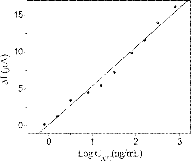
[0048] The preparation of the nanocomposite of embodiment 2 nano-platinum chitosan double coating thionine-graphene
[0049]Add 5 mg of thionine to 4.5 mL of graphene aqueous solution with a mass fraction of 0.05%, continue ultrasonication at 20° C. for 4 h, and centrifuge and wash with ethanol and deionized water, respectively, to remove thionine that is not combined with graphene, and obtain thionine- Graphene composite: Add the thionine-graphene composite to 5.5mL of 10mg / mL chitosan solution, stir continuously at 60°C for 12h, add 30μL of 0.1mol / L acetic acid dropwise, and stir at room temperature for 10min , continue heating in an oil bath at 90° C. for 1 h, and filter with a 0.26 μm nylon membrane to obtain thionine-graphene nanoparticles wrapped in chitosan; the thionine-graphene nanoparticles wrapped in chitosan Ultrasonic dispersion in water, take 5 mg and 120 μL of 1% HPtCl 6 Mix well and add to 10mL water, stir vigorously for 5min, add 0.25mL 100mmol / L NaBH dropwis...
Embodiment 3
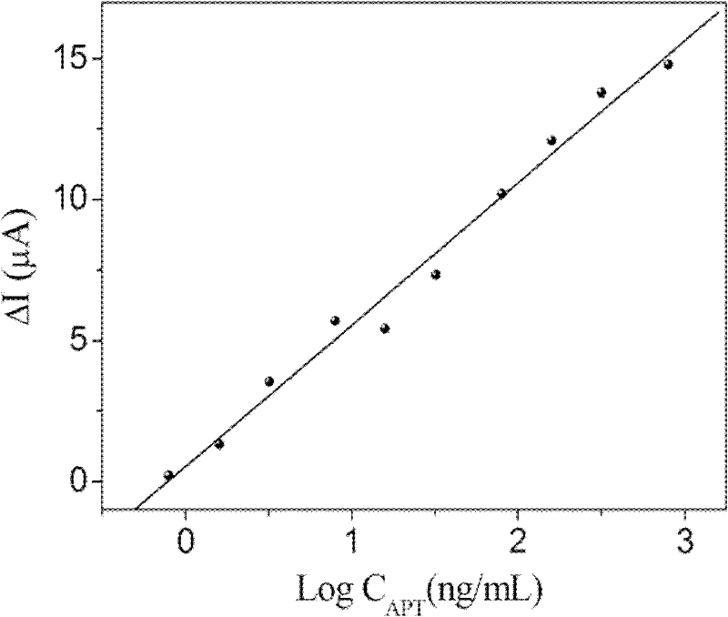
[0050] The preparation of the nanocomposite of embodiment 3 nano-platinum chitosan double coating thionine-graphene
[0051] Add 5 mg of thionine to 4.5 mL of graphene aqueous solution with a mass fraction of 0.05%, continue ultrasonication at 20° C. for 4 h, and centrifuge and wash with ethanol and deionized water, respectively, to remove thionine that is not combined with graphene, and obtain thionine- Graphene composite: Add the thionine-graphene composite to 5.5mL of 10mg / mL chitosan solution, stir continuously at 60°C for 12h, add 30μL of 0.1mol / L acetic acid dropwise, and stir at room temperature for 10min , continue heating in an oil bath at 90° C. for 1 h, and filter with a 0.22 μm nylon membrane to obtain thionine-graphene nanoparticles wrapped in chitosan; the thionine-graphene nanoparticles wrapped in chitosan Ultrasonic dispersion in water, take 5 mg and 150 μL of 1% HPtCl 6 Mix well and add to 10mL water, stir vigorously for 5min, add 0.25mL 100mmol / L NaBH dropwi...
PUM
 Abstract
Description
Claims
Application Information
 - R&D
- Intellectual Property
- Life Sciences
- Materials
- Tech Scout
- Unparalleled Data Quality
- Higher Quality Content
- 60% Fewer Hallucinations
Browse by: Latest US Patents, China's latest patents, Technical Efficacy Thesaurus, Application Domain, Technology Topic, Popular Technical Reports.
© 2025 PatSnap. All rights reserved.Legal|Privacy policy|Modern Slavery Act Transparency Statement|Sitemap|About US| Contact US: help@patsnap.com


