Quantitative analysis method based on lung MRI (magnetic resonance imaging) dynamic enhancement scanning

A dynamic enhancement and quantitative analysis technology, applied in the field of medical quantitative analysis, can solve the problems of measurement and calculation error, time-consuming calculation, hindering actual field application, etc., to achieve the effect of improving calculation efficiency and reducing work intensity

Inactive Publication Date: 2012-10-03
伍建林
View PDF2 Cites 6 Cited by
  • Summary
  • Abstract
  • Description
  • Claims
  • Application Information

AI Technical Summary

Problems solved by technology

[0004] Usually, the nuclear magnetic image signal is not linearly related to the change of contrast agent concentration in the tissue, so the use of non-model methods has great limitations. Currently, traditional model methods are mainly used for the quantitative analysis of lung DCE-MRI. The T1 algorithm is adopted. The calculation of T1(0) in the existing T1 algorithm mainly adopts the double flip angle method, which requires two scans of the patient, and for the lung scan, the tissue position will change every time the patient holds their breath. Larger errors are brought about; the lung tissue belongs to the dual blood supply system, and the ratio of tissue blood vessel volume is large, and the conventional model will produce larger errors when applied, which increases the complexity of the model; the blood supply of the lungs is complex, and in the traditional method Using a more complex model and AATT, the amount of calculation is quite large, especially when the tumor area is large, the calculation is very time-consuming; the MRI signal has large noise, which affects the accuracy of the calculation, and the point-by-point calculation will increase greatly The amount of calculations hinders the actual field application
In the method based on the pharmacokinetic model, it usually relies on the accurate measurement of the concentration change of the contrast agent in the blood vessel. However, due to the influence of time resolution, spatial resolution and respiratory motion, it is difficult to accurately measure the blood vessels around the lesion. Input function (AIF), these problems make quantitative analysis of lesions difficult to apply to clinical treatment

Method used


Image

  • Quantitative analysis method based on lung MRI (magnetic resonance imaging) dynamic enhancement scanning
  • Quantitative analysis method based on lung MRI (magnetic resonance imaging) dynamic enhancement scanning

Examples


Embodiment Construction

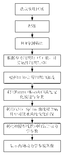
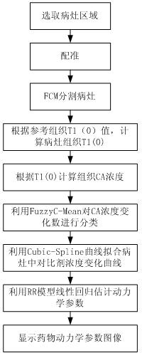
[0020] The reference region model (Reference Region Model) solves the need to directly measure the concentration change parameters (Artery Input Function) of the contrast agent in the artery. By measuring the concentration change of the reference region (usually the muscle tissue near the tumor), the The concentration change in the tumor area was used to obtain the relevant kinetic parameters. The Fuzzy C-Mean algorithm was introduced into DCE-MRI breast segmentation and pharmacokinetic classification. This method can classify clusters according to the change curve of the data, and can effectively solve the impact of noise on the data. The fixed T1 algorithm solves the conventional calculation of T1 images with multiple inversion angles, saves the number of scans of the patient, and greatly simplifies the calculation of T1 images. The present invention will combine the above several technologies and apply it to the quantitative analysis of DCE-MRI of the lungs. It adopts a fix...


PUM


Abstract

The invention relates to a quantitative analysis method based on lung MRI (magnetic resonance imaging) dynamic enhancement scanning. A reference region model, a FuzzyC-Mean algorithm and a fixed T1 algorithm are combined, and T1(0) of a tissue of interest is calculated through the T1 value of a given reference tissue, so that the shortcoming that scanning needs to be performed twice when a traditional double-flip angle Ti(0) calculation method is adopted can be overcome; a tissue model is taken as reference for avoiding the measurement of signals in an arterial region for getting an arterial input function, so that the working strength of researchers can be reduced; and automatic and precise segmentation is performed according to signal changes and the differences with a normal tissue in a tumor region during the period of injecting a contrast agent in the segmentation of a tumor in the lung, a traditional algorithm of calculating voxel by voxel in parameter estimation is changed, the voxels are classified according to the characteristics of concentration changes of the voxels, the calculation is performed gradually, and the calculation efficiency is greatly improved; and furthermore, the method has a better effect of suppressing the impacts of magnetic resonance signal noise on calculation result.

Description

technical field [0001] The present invention relates to a medical quantitative analysis technology, in particular to a quantitative analysis method applied to lung nuclear magnetic dynamic enhancement scanning (DCE-MRI) images, which is used to analyze the changes of drug hydrodynamic parameters in lung tumor tissue, and This is used to evaluate the effects of different stages of treatment. Background technique [0002] Lung cancer is currently the nauseating tumor with the highest morbidity and mortality. Its treatment has also become the focus of attention. Chemotherapy and radiotherapy are commonly used data methods. How doctors decide on late-stage treatment options plays a crucial role. [0003] In the existing technology, nuclear magnetic dynamic enhancement (DCE-MRI) method has been gradually applied to the evaluation of lung cancer treatment. This method mainly injects contrast agent into patients, analyzes the concentration change of contrast agent in the human bod...

Claims


Application Information

Patent Timeline
Patent Type & Authority Applications(China)
IPC IPC(8): G06F19/00
Inventor 伍建林张清苗延巍钟毅胡伽尼
Owner 伍建林
Why Eureka
Try Eureka