Preparation method for monoclonal antibody

A monoclonal antibody, antibody technology, applied in the fields of botanical equipment and methods, biochemical equipment and methods, chemical instruments and methods, etc., can solve the problems of high experimental costs and conditions, inability to use, etc.

Inactive Publication Date: 2012-11-14
BEIJING INST OF GENOMICS CHINESE ACAD OF SCI CHINA NAT CENT FOR BIOINFORMATION +1
View PDF0 Cites 9 Cited by
  • Summary
  • Abstract
  • Description
  • Claims
  • Application Information

AI Technical Summary

Problems solved by technology

This technology has the characteristics of high throughput and fast operation, and can complete the preparation of monoclonal antibodies within two weeks. However, the preparation of m

Method used


Image

  • Preparation method for monoclonal antibody
  • Preparation method for monoclonal antibody
  • Preparation method for monoclonal antibody

Examples


specific Embodiment 1

[0018] Preparation of Rabbit Lymphoid B Cells of Specific Example 1 Antigen Sensitization

[0019] The purified hbub3 protein was dissolved in 1×PBS, and an appropriate amount of the protein solution was mixed with complete Freund's adjuvant (Sigma) to emulsify. New Zealand big-eared white rabbits weighing 2Kg were taken, and the antigen was injected subcutaneously at multiple points on the back. After 3 weeks, take an appropriate amount of protein solution and mix and emulsify it with Freund's incomplete adjuvant (Sigma), and carry out the second immunization subcutaneously on the back of the big-eared white rabbit, and carry out the third immunization every 3 weeks. -7 days, take peripheral blood lymphocytes.

specific Embodiment 2

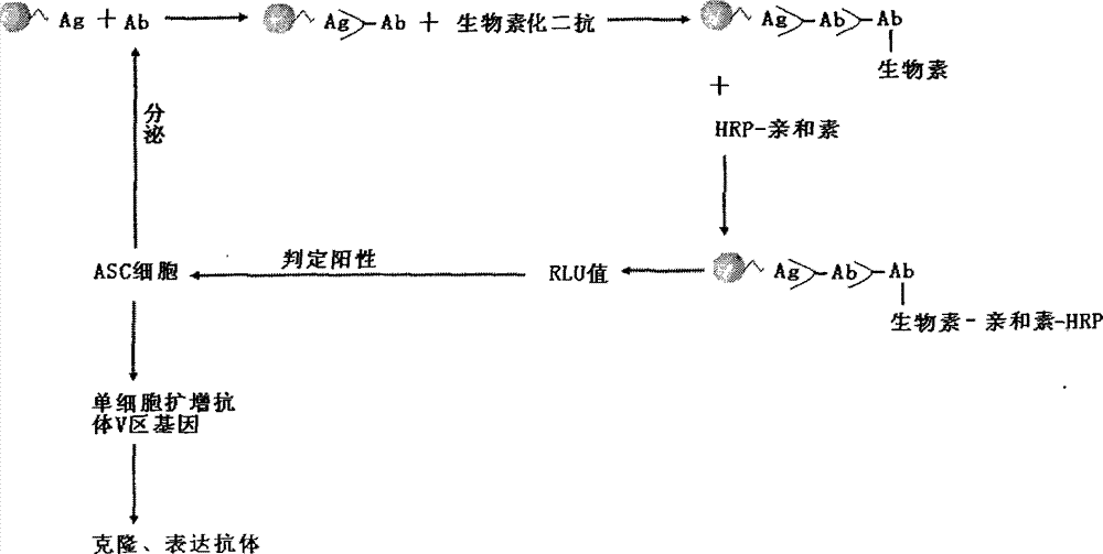
[0020] Specific Example 2 Screening of Antibody Secretion Positive Cells

[0021] (1) Antigen coupling

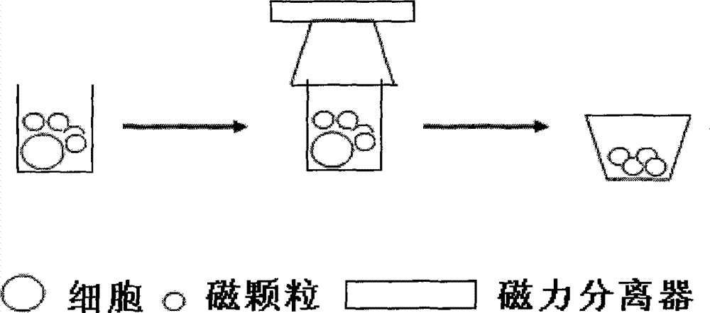
[0022] Take 3 mg of dry powder of magnetic particles with a diameter of 2.8 μm and epoxy groups on the surface (Dynabeads M-270Epoxy, Invitrogen Company), add 200 μl of 0.1M PB, pH 7.4, oscillate and suspend, rest for 10 minutes, and place on the magnetic stand for 2 minutes Aggregate the magnetic particles, remove the liquid, repeat washing twice and add. Take 60 μl volume of 1mg hbub3 antigen (soluble antigen is dissolved in PB, inclusion body is redissolved in SDS, SDS final concentration is 1%), add 60 μl 0.1M PB, pH 7.4 suspended magnetic particles, shake and mix. Then add 60μl 3M(NH4) to the tube 2 SO 4(dissolved in 0.1M PB, pH 7.4), mix well, and rotate for 20 hours at 37°C. After the reaction, put it on the magnetic stand for 4 minutes, separate the magnetic particles, and wash twice with 0.1% BSAPBS.

[0023] (2) Antibody detection

[0024] Take peripheral bl...

specific Embodiment 3

[0025] Specific Example 3 Antibody Variable Region Gene Amplification

[0026] Move the cell culture plate from -80°C to room temperature (the following operations are performed according to RNA operation), transfer the cell lysate in the positive well to the RT-PCR tube, and add 0.5 μl RNasin. Using the primers located in the antibody constant region as specific primers, SuperScript III First-Strand Synthesis (Invitrogen) was used to synthesize antibody heavy chain and light chain cDNA. The above primers located at the 5'-end Leader and FRI regions are used as upstream primers, and the primers located in the antibody constant region are used as downstream primers to perform nest PCR to amplify the antibody heavy chain and light chain variable region genes. The RT primer for amplifying the heavy chain of the rabbit antibody is located in the constant region of the antibody, the sequence is as shown in SEQ ID NO: 1, the sequence of the upstream primer of the first PCR is as SEQ...


PUM


Abstract

The invention discloses a preparation method for a monoclonal antibody. According to the method, magnetic granular microballoon spheres of an immobilized antigen are added into B lymphocyte culture micropores, magnetic separation is carried out on the magnetic granular microballoon spheres so as to allow the spheres to enter into corresponding microplates, a single B lymphocyte which secretes a specific antibody is detected by using the method of immunochemiluminescence or immunofluorescence, a light chain variable region gene and a heavy chain variable region gene of the antibody are amplified by using the method of single-cell RT-PCR and nested RT-PCR and are recombined to expression plasmid containing a constant domain of the antibody, and a host cell is transfected to express the monoclonal antibody.

Description

technical field [0001] The invention relates to the technical fields of bioengineering and monoclonal antibody production. Specifically, the present invention relates to the detection and recognition of a single B cell that secretes an antigen-specific antibody, the antibody light and heavy chain variable region genes are amplified by RT-PCR and PCR methods, and constructed on a vector containing the constant region gene through homologous recombination , Transfected cells to prepare monoclonal antibodies. Background technique [0002] Antibodies are produced by the body's immune system under the stimulation of antigens by the synthesis and secretion of plasma cells that proliferate and differentiate from B lymphocytes or memory cells. They are immunoglobulins that can specifically bind to corresponding antigens and are important in mediating humoral immunity. immune effector molecules. Antibodies have the functions of neutralizing viruses, activating the complement system...

Claims


Application Information

Patent Timeline
IPC IPC(8): C12N15/13C12N15/63C12N15/11C07K16/00
Inventor 邵长君高静王绪敏冯杰于军
Owner BEIJING INST OF GENOMICS CHINESE ACAD OF SCI CHINA NAT CENT FOR BIOINFORMATION
Why Eureka
Try Eureka