Specific interaction of dentritic cell factor 1 (DCF1) genes and transmembrane protein 59 like (TMEM 59 L) genes
A TMEM59L, interaction technology, applied in recombinant DNA technology, microbial assay/inspection, biochemical equipment and methods, etc., can solve problems such as limited research
- Summary
- Abstract
- Description
- Claims
- Application Information
AI Technical Summary
Problems solved by technology
Method used
Image
Examples
Embodiment 1
[0017] Embodiment 1: vector construction
[0018]Use Olige 6.0 software to design primers for human DCF1, mouse DCF1, human TMEM59L, mouse TMEM59L, APP(695), GDI1, GDI2, and BACE2 genes, and introduce restriction sites and protective bases into the upstream and downstream primers, respectively. After the genes were amplified by PCR, they were connected into the corresponding vectors, as shown in Table 1.
[0019]
Embodiment 2
[0020] Example 2: Plasmid transfected cells
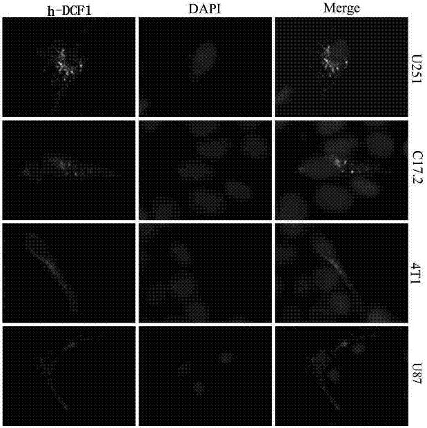
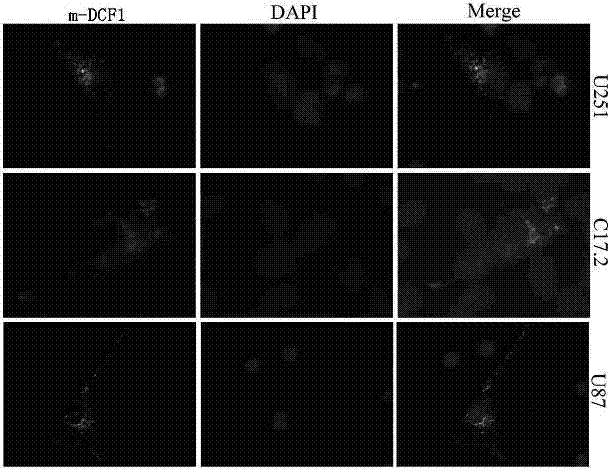
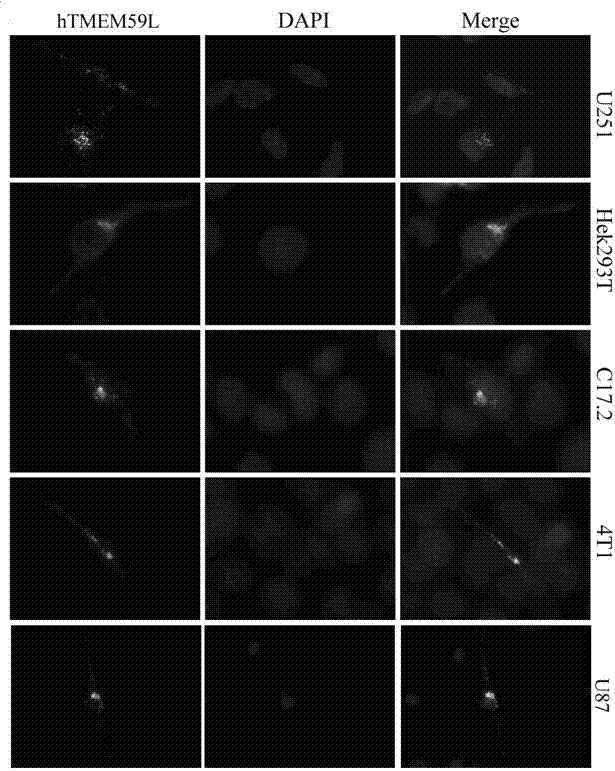
[0021] Transfect human and mouse DCF1 and TMEM59L plasmids into U251, HEK293T, C17.2, 4T1, U87 and other cells. The transfection process is as follows:
[0022] 1. The day before transfection, cells were trypsinized and counted, and the cells were plated in a 6-well plate so that the density on the day of transfection was 70%. Add 1.5mL of serum-containing, normal growth medium without antibiotics to each well.
[0023] 2. When transfecting, first replace the serum-containing medium in the 6-well plate with serum-free medium, 1 mL per well, and place at 37°C, 5% CO 2 in the incubator.
[0024] 3. For each well of cells, dilute 3 μg of plasmid DNA with 250 μL of serum-free medium DMEM.
[0025] 4. For each well of cells, dilute 3 μL Lipofectamine 2000 reagent with 250 μL serum-free medium DMEM, and let stand at room temperature for 5 minutes.
[0026] 5. Mix diluted plasmid DNA (step 3) and diluted Lipofectamine 2000 (st...
Embodiment 3
[0031] Example Three: Organelle Staining
[0032] After DCF1 and TMEM59L were transfected with green or red fluorescent plasmids, they were stained with corresponding organelle dyes for co-localization observation.
[0033] (1) Fluorescent labeling of the Golgi apparatus
[0034] Remove the cell culture medium, and wash the cells grown on the coverslip with an appropriate amount of solution such as D-Hanks solution. Remove the washing solution, add the prepared Golgi-Tracker Red staining solution, and incubate with the cells at 4°C for 30 minutes. Recover the Golgi-Tracker Red staining working solution, wash the cells about 3 times with the cell culture medium pre-cooled in an ice bath, replace with fresh culture medium and incubate at 37°C for 30 minutes. After washing once more with fresh culture medium, observation is usually performed with a fluorescence microscope or a confocal laser microscope. At this time, it can be observed that the Golgi apparatus shows bright a...
PUM
 Abstract
Description
Claims
Application Information
 - R&D
- Intellectual Property
- Life Sciences
- Materials
- Tech Scout
- Unparalleled Data Quality
- Higher Quality Content
- 60% Fewer Hallucinations
Browse by: Latest US Patents, China's latest patents, Technical Efficacy Thesaurus, Application Domain, Technology Topic, Popular Technical Reports.
© 2025 PatSnap. All rights reserved.Legal|Privacy policy|Modern Slavery Act Transparency Statement|Sitemap|About US| Contact US: help@patsnap.com


