
Multifunctional biomedical microscope

A biomedical and multifunctional technology, applied in the field of multifunctional biomedical microscopy, can solve the problems of shallow tomographic depth and low lateral and axial resolution of a single optical coherence tomography system, and overcome the problem of shallow tomographic depth Effect

Inactive Publication Date: 2012-12-12
SUZHOU MICROCLEAR MEDICAL INSTR
View PDF7 Cites 22 Cited by
  • Summary
  • Abstract
  • Description
  • Claims
  • Application Information

AI Technical Summary

Problems solved by technology

[0007]1) Overcome the shortcoming of shallow tomographic depth of a single laser confocal microscope; 2) Overcome the shortcoming of low lateral and axial resolution of a single optical coherence tomography system

Method used


Image

  • Multifunctional biomedical microscope
  • Multifunctional biomedical microscope

Examples


Embodiment Construction

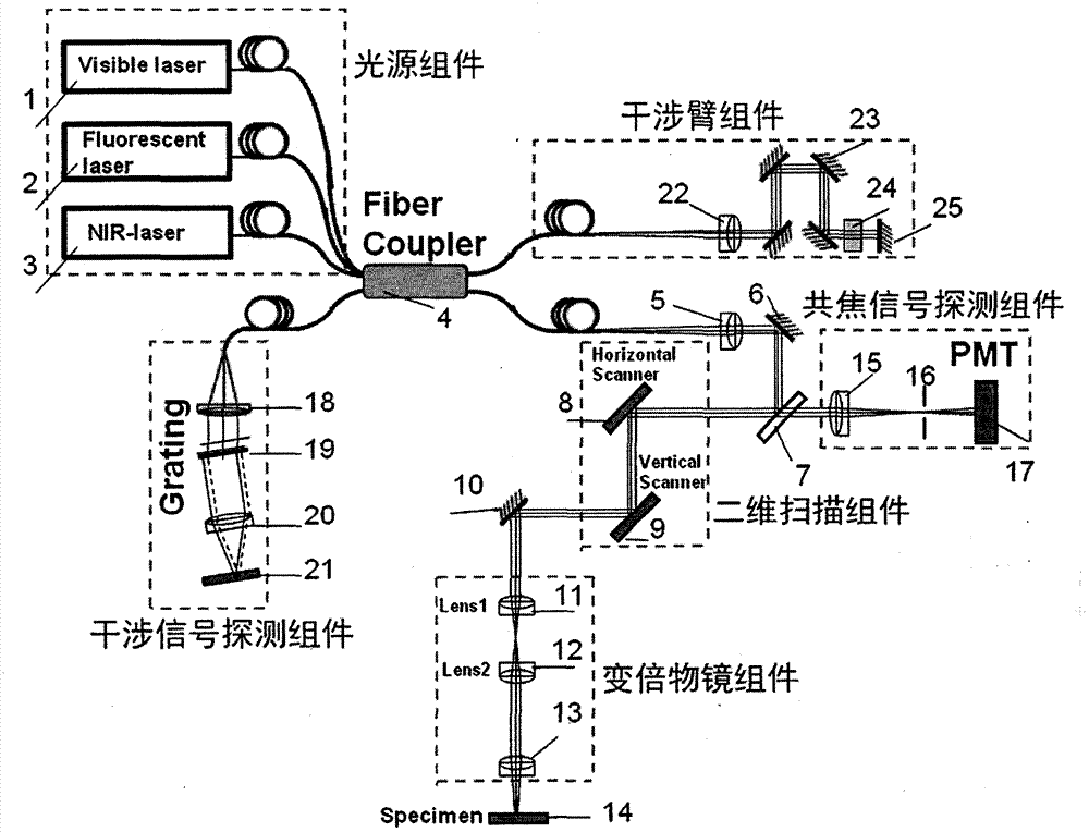
[0015] According to the instructions attached figure 1 , How to specifically implement the functions of the biomedical microscope proposed in the present invention is described in detail as follows:

[0016] 1. The illumination beam generated by the visible light source (1) of the light source assembly enters the fiber coupling lens (5) through one of the two output ends of the multi-path fiber coupler (4), and is reflected by the mirror (6) in turn After splitting light with the beam splitter (7), it enters the two-dimensional scanning assembly (8, 9). The illuminating laser beams emitted from the two-dimensional scanning components (8, 9) are sequentially reflected by the reflector (10), and focused by the variable magnification objective lens component (11-13) to illuminate the sample (14) to be tested.

[0017] 2. The visible light source of the light source assembly (1) Generally, it is a semiconductor laser, which is used to generate a laser beam in the visible light band. Th...


PUM


Abstract

The invention relates to a multifunctional biomedical microscope with the functions of laser scanning confocal imaging, fluorescence imaging and optical coherence tomography. The multifunctional biomedical microscope is composed of a light source component, a two-dimensional scanning component, a zoom objective component, a confocal signal detecting component, an interference arm component and an interference signal detecting component. According to the multifunctional biomedical microscope, a confocal image of a sample is obtained by the introduction of a visible-light source lighting sample, a fluorescence image of the sample is obtained by the introduction of a fluorescence light source lighting sample, and an optical coherence tomography image of the sample is obtained by the introduction of a broadband low-coherence near-infrared laser light source lighting sample, and the multifunctional biomedical microscope is realized so as to obtain a micro-scale three-dimensional organization structure image of a transparent or non-transparent sample.

Description

technical field [0001] The invention relates to a multifunctional biomedical microscope, in particular to a multifunctional biomedical microscope used for biomedical tissue imaging and capable of obtaining confocal images, fluorescence images and optical coherence tomography images. Background technique [0002] The concept of confocal microscopy was first proposed by Minsky in the United States in the late 1950s (Marvin Minsky. "Memo iron inventing confocal scanning microscope", Scanning, 1988, 128-138), and then P.Davidovits , A.F.Slomba and C.J.R.Sheppard and many other scholars have conducted more detailed research on confocal imaging. Confocal laser microscopy systems, especially confocal fluorescence microscopy systems combined with fluorescent probe detection technology, have higher lateral resolution (about 100nm) and longitudinal resolution due to the use of detection pinholes and shorter wavelength lasers. The rate (about 50nm) is widely used in the detection of b...

Claims


Application Information

Patent Timeline
IPC IPC(8): G01N21/00G01N21/64
Inventor 李超宏杨丛渊张锐进
Owner SUZHOU MICROCLEAR MEDICAL INSTR
Features
  • Generate Ideas
  • Intellectual Property
  • Life Sciences
  • Materials
  • Tech Scout
  • Unparalleled Data Quality
  • Higher Quality Content
  • 60% Fewer Hallucinations
Learn More