
Capsule endoscope feeding device

A technology for capsule endoscope and gastroscope, which is applied in the field of medical appliances, can solve the problems of limited battery life, poor device reliability, and inability to cut off, and achieves the effects of low manufacturing cost, compact structure, and reliable release.

Inactive Publication Date: 2013-01-16
WEST CHINA HOSPITAL SICHUAN UNIV
View PDF9 Cites 5 Cited by
  • Summary
  • Abstract
  • Description
  • Claims
  • Application Information

AI Technical Summary

Problems solved by technology

Although capsule endoscopy has high diagnostic value for diseases such as small intestinal inflammation and bleeding, it also has many defects. One of the more common shortcomings is that capsule endoscopy is greatly affected by the movement (peristalsis) of the stomach and small intestine after swallowing, while Battery life is limited
When the peristalsis of the stomach or esophagus is slow or there is obstruction, the capsule will stay in the stomach or esophagus for a long time, and cannot enter the small intestine or the battery energy has been consumed too much when entering the small intestine, and it is no longer possible to take pictures of the entire small intestine. Missed diagnosis due to inability to complete the examination
Another less common disadvantage is that capsule endoscopy is still too bulky compared to clinically administered capsule-shaped pills, and very few patients (mainly young patients) cannot swallow due to fear or throat muscle spasm. Into the esophagus, making the test impossible
[0003] When the above situation occurs, it is necessary for the physician to send the capsule endoscope into the descending duodenum with the help of gastroscope and endoscope snare (or basket), which increases the pain of the patient, is time-consuming, labor-intensive, and may unsuccessful
In addition, capsule endoscopy is currently very expensive. Once the inspection is unsuccessful, it will easily lead to conflicts among patients, hospitals, distributors and manufacturers.
[0004] In view of the above problems, relevant patent literature has reported that a special pusher can be used to deliver the capsule endoscope to the duodenum with the assistance of a gastroscope. It is more complicated, and it is only for patients who cannot swallow capsule endoscopes smoothly. As mentioned above, the shell of capsule endoscopes is made of smooth polymer materials. In clinical practice, most patients can swallow them smoothly and enter the 12 the duodenum, so in adults, the routine use of a pusher to enter the duodenum is not required
However, the shell of the capsule endoscope is very smooth, and it is even more difficult to take the retained capsule endoscope out of the body through the gastroscope than directly into the duodenum, so the practical value of the pusher is very limited
At present, clinically, for capsule endoscopy that stays in the esophagus and stomach, under the guidance of a gastroscope, a snare or a basket is used to trap the capsule and send it to the duodenum. Smooth, the snare or basket is not easy to net the capsule, even after the net is caught, it can only net the middle part of the capsule endoscope shell horizontally. The resistance to passing through the pylorus is very high, and the steel wire of the snare or the basket can not resist the resistance and bend, resulting in the failure of the capsule endoscope to pass through the pylorus or slippage. Therefore, gastroscopy doctors need to spend a lot of time and energy to complete this work, and the pressure is very high during the operation. At the same time, the gastroscope stays in the patient's body for a long time, which increases the pain and risk of the patient
[0005] CN200480028555.1 discloses a digestive canal inspection device, including a capsule endoscope capable of digestive canal inspection, a flexible tubular body, and a flexible rope-like member inserted into the tubular body. A connecting part is provided in the capsule endoscope, and the rope-shaped part is detachably connected to the connecting part of the capsule endoscope to detachably connect the tube body and the capsule endoscope. It is necessary to make the connection on the capsule endoscope The part makes the structure complicated and needs to be specially made, which increases the production cost and affects the smoothness of the surface of the capsule endoscope. When disassembling and separating, it is necessary to cut the connecting rope or destroy the connecting part, which may occur when there is no need for separation. The connecting rope is cut or the connecting part is damaged, and it may not be possible to cut the connecting rope or destroy the connecting part when separation is required. Therefore, the reliability of the device is poor and the operation is inconvenient
Also, capsule endoscopes lodged in the stomach or digestive tract cannot be picked up with this device

Method used


Image

  • Capsule endoscope feeding device
  • Capsule endoscope feeding device
  • Capsule endoscope feeding device

Examples


Embodiment Construction

[0021] The present invention will be further described below in conjunction with the accompanying drawings.

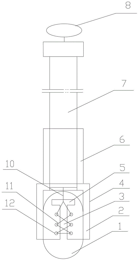
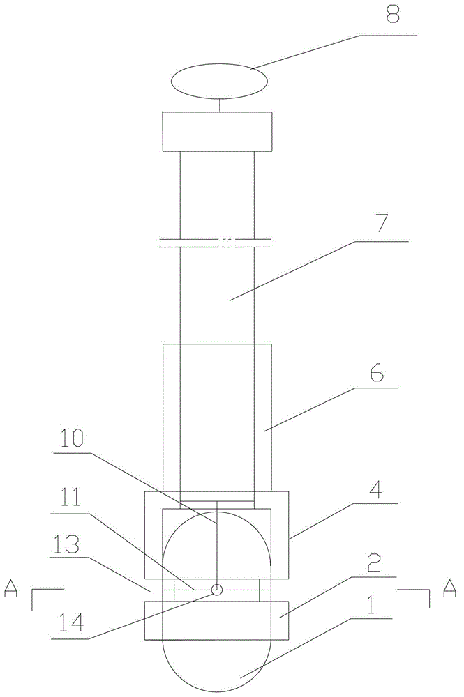
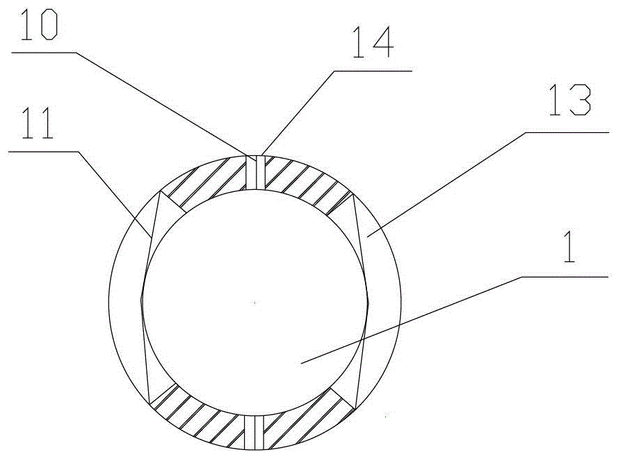
[0022] As shown in the figure, the capsule endoscope feeder of the present invention includes a gastroscope tube 7 , a connecting sleeve 4 , a fixing line 11 , and a control line 10 . The small end 6 of the connecting sleeve 4 is mounted on the front end of the gastroscope tube 7, the inner diameter of the large end 2 is slightly larger than the outer diameter of the capsule endoscope 1, and the length is 2 / 3 of the length of the capsule endoscope 1, and the capsule endoscope 1 can be installed on the large end 2; the fixed line 11 is installed on the large end 2 of the connecting sleeve for fixing the capsule endoscope 1, and the control line 10 passes through the gastroscope tube 7 and is connected to the fixed line 11.

[0023] Preferably, the connecting sleeve 4 is made of elastic transparent medical material, such as plastic.

[0024] Further, such as figure 1 A...


PUM


Abstract

A capsule endoscope feeding device comprises a gastroscope tube, a connection sleeve, a fixing wire and a control wire, wherein the small end of the connection sleeve is arranged on the gastroscope tube, the inner diameter of the larger end is slightly larger than the outer diameter of a capsule endoscope, the length of the connection sleeve is two thirds of the length of the capsule endoscope, and the capsule endoscope is arranged in the large end; the fixing wire is arranged on the large end of the connection sleeve to fix the capsule endoscope; and the control wire penetrates through the gastroscope tube to be connected with the fixing wire. According to the capsule endoscope feeding device, the capsule endoscope left in the alimentary canal or the stomach can be conveniently and quickly taken and fed into the duodenum; for a patient having difficulty in swallowing the capsule endoscope, the capsule endoscope outside the body can be quickly fed into the duodenum; the capsule endoscope feeding device can release the capsule endoscope reliably, is compact in structure and low in manufacture cost, and can be used for the existing universal capsule endoscope.

Description

technical field [0001] The invention relates to a medical appliance, in particular to a capsule endoscope feeder for digestive tract inspection. Background technique [0002] The small intestine is the lumen of the digestive tract connecting the pylorus and the ileocecal valve. Except for the part above the descending duodenum and the terminal ileum, which can be subjected to routine gastroscopy and colonoscopy, the other parts are the most difficult to examine in the entire gastrointestinal tract. Due to the long length of the small intestine (3.35-7.85m), the free intraperitoneum and the formation of multiple compound intestinal loops bound by the mesentery, the traditional inspection techniques are greatly limited. Many methods of small intestine examination, such as X-ray small intestine barium meal examination, enteroscopy, radionuclide scanning, and arteriography, all have low diagnostic positive rate, inaccurate positioning and qualitative identification, great pain f...

Claims


Application Information

Patent Timeline
IPC IPC(8): A61B1/00A61B5/07
Inventor 甘涛吴俊超王一平
Owner WEST CHINA HOSPITAL SICHUAN UNIV
Learn More