

A tissue cryopreservation solution that can maintain cell viability
A technique for cryopreservation and tissue block, which is applied in the biological field
- Summary
- Abstract
- Description
- Claims
- Application Information
AI Technical Summary
Problems solved by technology
Method used
Image

Examples
Embodiment 1
[0087] cryopreservation of adipose tissue
[0088] Experimental Materials
[0089] Adipose tissue: take 20ml of fresh sterile adipose tissue from the operating room
[0090] Freezing solution: KSR solution of 80vol%KSR+10vol%DMSO+10vol% trehalose, wherein the concentration of the KSR solution of trehalose is 2mol / l.
[0091] Experimental group
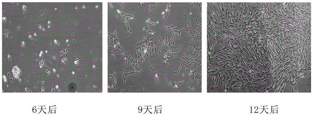
[0092] Panel A: cells were extracted from fresh adipose tissue and cultured
[0093] Group B: cells were extracted from frozen adipose tissue and cultured
[0094] experimental method
[0095] (1) Processing of fresh adipose tissue
[0096] Place the fresh adipose tissue in a sterile bottle under aseptic conditions, and wash with DPBS or saline for 3-4 times to remove the miscellaneous cells on the surface of the adipose tissue. Then use sterilized scissors to cut the tissue pieces (approximately 1mm 3 size) to facilitate cryopreservation of tissues.
[0097] The shredded tissues were equally divided into two groups, group A and ...
Embodiment 2
[0105] Recovery and inoculation of adipose tissue
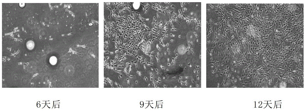
[0106] After 3 months, the cryopreservation tube containing the frozen adipose tissue was quickly placed in a 37°C water bath. After the tissue was dissolved, the tissue was taken out, and the residual cryopreservation solution was washed with DMEM (Sigma Company). Inoculate the resuscitated tissue into a culture flask of the same size and specification as Group A, add culture medium, and place at 37°C, CO 2 cultured in an incubator. Tissues were inoculated, and the tissue was completely changed after 3 days. The adherent growth of cells was observed under a microscope. Also make a record every 3 days and take pictures. The result is as figure 2 shown. After 3 days, no adherent cells were found; after 6 days, very few adherent cells were found, and the adherent cells were long spindle-shaped; after 9 days, the amount of adherent cells increased, and the cells grew well; after 12 days, there were many adherent cells, and...
Embodiment 3
[0113] Flow cytometry detection
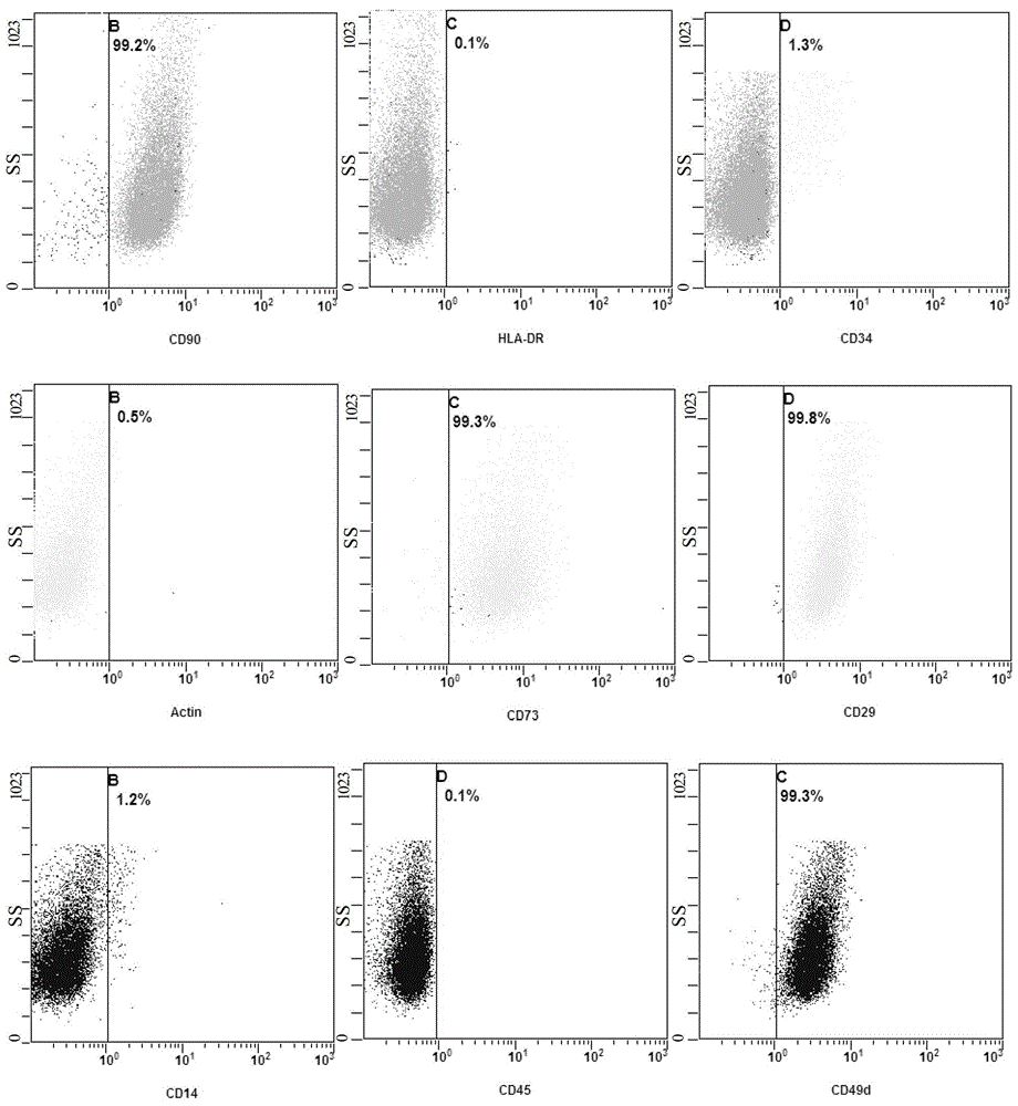
[0114] The above-mentioned cryopreserved P3 generation cells were taken, and after thawing, they were made into a cell suspension, and flow cytometric detection was performed to determine that the obtained cells were adipose-derived mesenchymal stem cells, and the cells were identified in terms of cell purity. Specifically, the cells were taken to make a cell suspension, and the density was adjusted to 1×10 5 mL -1 , Centrifuge at 800r / min (120g) for 5min, discard the supernatant, wash the resuspended cells with cold D-Hanks at 4°C, centrifuge the cell suspension again at 800r / min for 5min, and discard the supernatant. Then the cells were resuspended to 1 mL with D-Hanks, antibodies were added, and cell surface markers were detected by flow cytometry. The added antibodies were: human anti-CD29, CD73, CD49d, CD90, CD14, CD45, CD34, Actin and HLA-DR. The result is as image 3 and shown in Table 2. Among them, CD29, CD73, CD49d, and CD90 are...
PUM
 Abstract
Description
Claims
Application Information