Establishment and characterization method of in-vitro blood brain barrier model based on microfluidic chip

A microfluidic chip and blood-brain barrier technology, applied in biochemical equipment and methods, artificial cell constructs, tissue cell/virus culture devices, etc., can solve difficult blood-brain barrier models and difficult to achieve mesenchymal stem cells In-situ observation and other issues to achieve the effect of reducing consumption

Inactive Publication Date: 2013-03-20
DALIAN INST OF CHEM PHYSICS CHINESE ACAD OF SCI
View PDF4 Cites 37 Cited by
  • Summary
  • Abstract
  • Description
  • Claims
  • Application Information

AI Technical Summary

Problems solved by technology

Existing in vitro blood-brain barrier simulation methods mainly focus on commercial orifice plates or Transwell chambers to study the structure and function of two-dimensional blood-brain barrier models. The main problem is that orifice plates or Transwell chambers are difficult to construct in vitro There is a blood-brain barrier model that transitions from a two-dimensional plane to a three-...

Method used


Image

  • Establishment and characterization method of in-vitro blood brain barrier model based on microfluidic chip
  • Establishment and characterization method of in-vitro blood brain barrier model based on microfluidic chip
  • Establishment and characterization method of in-vitro blood brain barrier model based on microfluidic chip

Examples


Embodiment 1

[0032] HUVEC cells tightly fused on a three-dimensional collagen surface to form the blood-brain barrier and its structural characterization

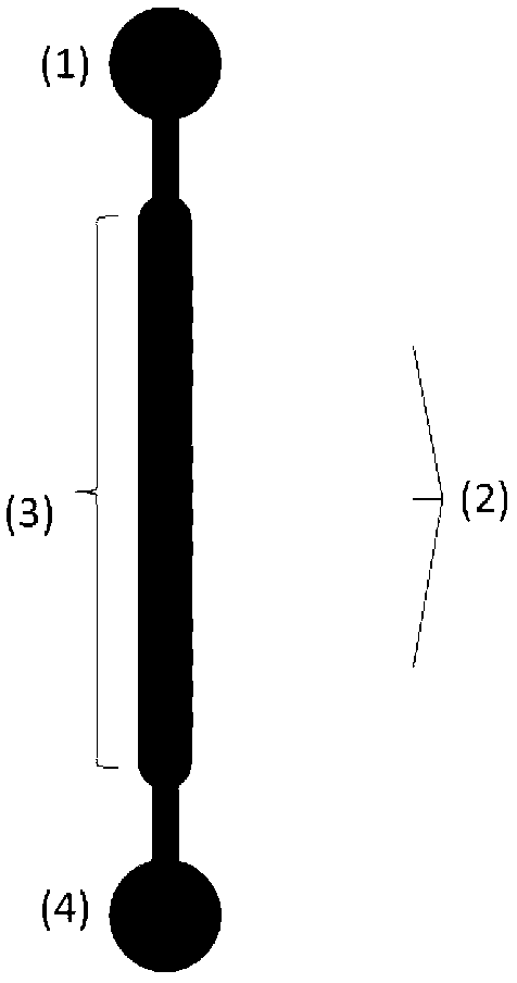
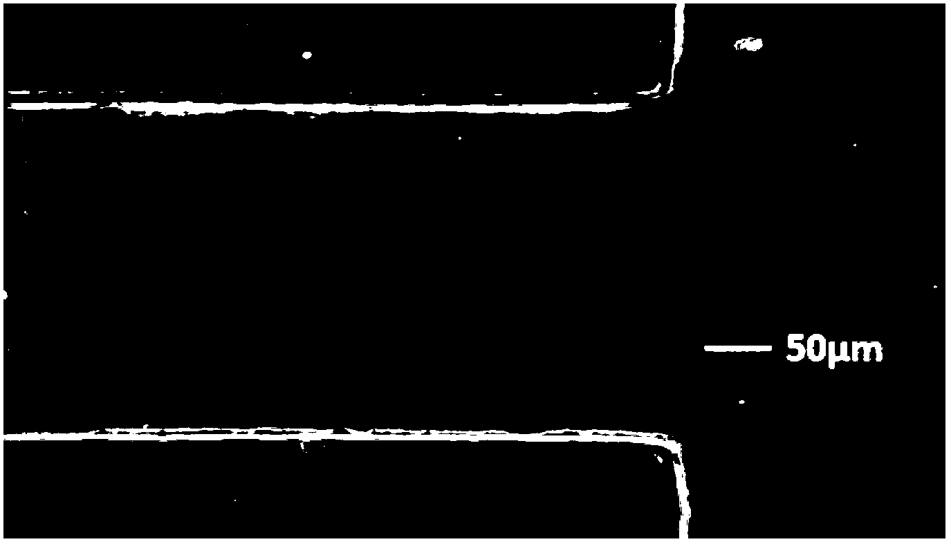
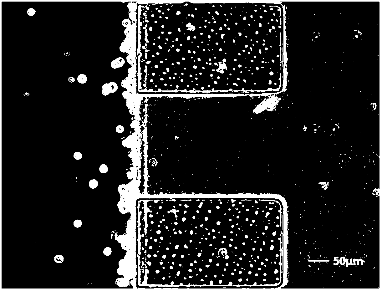
[0033] Using the microfluidic chip designed and manufactured by the laboratory, the configuration is as follows: figure 1 shown. Prepare a collagen stock solution with a concentration of 3 mg / mL, add 0.4 μL per well into the collagen inlet pool, gel at 37 °C for 30 min, and the distribution of the formed glue filaments is as follows: figure 2 shown. Add 10-20 μL of HUVEC cell suspension from the cell inlet pool, and the concentration of the cell suspension is 5×10 6 -1×10 7 cells / mL, the chip was erected for 10 minutes to make the cells attach to the collagen side, such as image 3 As shown, take pictures to record the initial position of the cells. Change the medium every 24h, and take pictures to record the fusion of HUVEC cells, such as Figure 4 shown. After 48 hours, cell immunofluorescence staining was carried out, and the...

Embodiment 2

[0035] Characterization of Blood Brain Barrier Permeability Using Small Molecule Fluorescent Substances

[0036] Using the microfluidic chip designed and manufactured by the laboratory, the configuration is as follows: figure 1shown. After the 3 mg / mL collagen was perfused into the chip and coagulated, the blood-brain barrier model was established using the same cell inoculation and culture methods as in Example 1. 48 hours after HUVEC cells were inoculated into the chip, 10μLCellTracker Green was added to the chip with a concentration of 10μmol / L and a molecular weight of 557.47D. From 0min, the fluorescence intensity on the side of the three-dimensional collagen was recorded every 15min, and statistical calculation was performed to characterize the permeability of the blood-brain barrier. The result is as Figure 6 shown.


PUM


Abstract

The present invention provides an establishment and characterization method of in-vitro blood brain barrier model based on microfluidic chip. The microfluidic chip mainly comprises a cell inlet pool (1), collagen inlet pools (2), a cell culture chamber (3) and a waste liquid pool (4). The upper of the cell culture chamber (3) is connected with the cell inlet pool (1). The lower of the cell culture chamber (3) is connected with the waste liquid pool (4). Each collagen inlet pool respectively includes four observation chambers. The collagen inlet pools are communicated with the cell culture chamber (3). According to the invention, construction and characterization of the in-vitro blood brain barrier model and evaluation of the barrier function are integrated into a chip of a few square centimeters, and the chip can be used for in-vitro simulation and subsequent applications of the blood-brain barrier model. Compared the present invention with a Transwell small chamber co-culture model, the cell co-culture model secondary vaccination and time-consuming problems are solved, the flow conditions are added, the chip is closer to the true in-vivo microenvironment, the cell and reagent consumption are significantly reduced, and a plurality of experiment parameters can be obtained once and simultaneously.

Description

technical field [0001] The invention relates to the field of simulation and application of applying microfluidic chip technology to in vivo tissue engineering, in particular to a method for establishing and characterizing an in vitro blood-brain barrier model based on a microfluidic chip. Background technique [0002] In 1885, German Paul Ehrlich firstly proved that the permeability of cerebral blood vessels is different from that of non-nervous blood vessels. Later researchers successively realized that there is a barrier between blood and brain, and called it the blood-brain barrier. The blood-brain barrier is a structural and functional barrier composed of brain microvascular endothelial cells, astrocytes, and pericytes, which regulates the flow of molecules into and out of the brain to maintain the neural microenvironment. Many diseases in the brain are related to the destruction of the defense function of the blood-brain barrier, and drugs cannot pass through the blood-...

Claims


Application Information

Patent Timeline
IPC IPC(8): C12M3/00C12N5/071
Inventor 秦建华许慧马慧朋
Owner DALIAN INST OF CHEM PHYSICS CHINESE ACAD OF SCI
Why Eureka
Try Eureka