Immunoliposome biochip, preparation method thereof and application thereof in biological detection

A technology of immunoliposome and plastid biology, which is applied in the direction of measuring devices, instruments, scientific instruments, etc., can solve the problems of unsuitable high-throughput analysis and time-consuming, and achieve the effect of reducing dosage and improving binding ability

Active Publication Date: 2013-07-10
美国纳米材料创新有限公司 +1
View PDF10 Cites 22 Cited by
  • Summary
  • Abstract
  • Description
  • Claims
  • Application Information

AI Technical Summary

Problems solved by technology

The isolation and characterization of microRNA in exosomes involves a series of centrifugation steps, usually by ultracentrifugation on a sucrose gradient, but it is difficult to obtain high-purity exosomes using this method, and this method is time-consuming and not suitable for High-throughput analysis in the clinic

Method used


Image

  • Immunoliposome biochip, preparation method thereof and application thereof in biological detection
  • Immunoliposome biochip, preparation method thereof and application thereof in biological detection
  • Immunoliposome biochip, preparation method thereof and application thereof in biological detection

Examples


Embodiment 1

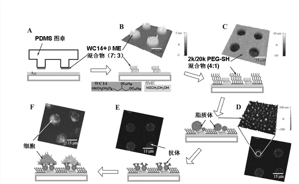
[0038] Example 1: Fabrication of immunoliposome (tILN) biochip

[0039] Example 1 describes a simple method for fabrication of anchored immunoliposome biochips. Such as figure 1 As shown, using a PDMS stamp, an ethanol solution containing 7:3 (molar ratio) of WC14 and β-mercaptoethanol (βME) was transferred to a gold-coated glass substrate by microcontact printing, and the PDMS stamp was fabricated using soft etching techniques. In an atomic force microscope (AFM) image, figure 1 B shows a chip with a diameter of 15 μm, the height of the patterned area is about 4 nm.

[0040] To prevent non-specific cell binding, we invented a mixture of PEG-SH with a molecular weight of 2,000 and 20,000 Daltons in a 4:1 molar ratio to protect 80% of the non-microarray area. In the AFM image, figure 1 The height of PEG-SH shown in C is about 10 nm. Such as figure 1 As shown in D, 10 mg / ml lipid mixture [egg phosphatidylcholine (Egg PC): cholesterol (CHOL): distearoylphosphatidylethanola...

Embodiment 2

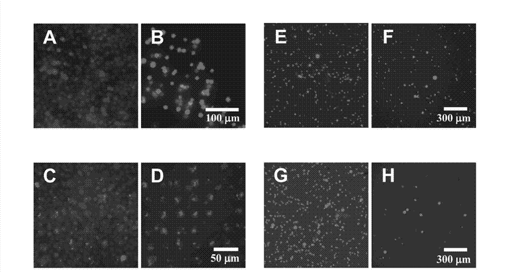
[0042] Example 2: Isolation of Raji cells from Jurkat cells using an anti-CD20-based chip

[0043] Example 2 involved the isolation of Raji cells (Burkitt lymphoma B cell line) from Jurkat cells (acute lymphoblastic leukemia cell line) using a cell surface ligand chip containing Rituximab (anti-CD20 monoclonal antibody rituximab). figure 2 Among them, (A) to (D) fluorescence microscope images show the comparison of the results of capturing cells with conventional antibodies and the immunoliposome biochip prepared in Example 1. Apparently, the effect of isolating Raji cells with immunoliposome biochip is much better.

Embodiment 3

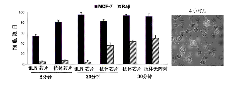
[0044] Example 3: Isolation of MCF-7 cells from Raji cells with EpCAM antibody-based chip

[0045] Example 3 is the application of the immunoliposome biochip prepared in Example 1 to the isolation of rare cells, such as circulating tumor cells (CTCs) in blood samples, which are very valuable for early clinical diagnosis and non-invasive cancer metastasis Prognostic indicators. However, it is still a huge challenge to detect CTCs in medicine, mainly because the number of such cells in the blood of patients is very small. Since CTCs express epithelial cell adhesion molecule (EpCAM) on their surface, but normal blood cells do not, immobilized anti-EpCAM has been used on CTCs isolated from patient blood. In this example, our target simulates blood cells to separate a very small amount of MCF-7 cells from a large number of Raji cells. At the same time, because the overexpression of miR-21 in breast cancer cells can be used as a viable biomarker. Using these cell lines at differe...


PUM

PropertyMeasurementUnit
particle diameteraaaaaaaaaa
diameteraaaaaaaaaa

Abstract

The invention relates to an immunoliposome biochip, a preparation method thereof and an application thereof in biological detection. The immunoliposome provided by the invention is distributed on the chip in a micro-array way, and includes molecule probes, such as molecular beacon with fluorescence; and outside of the immunoliposome is loaded with various ligands. The immunoliposome biochip can specifically capture target cells in a cell mixture, and ensure messenger RNA and/or micromolecule RNA in the target cells without destroying biomarkers in the cells. In addition, the immunoliposome biochip provided by the invention can be used for capturing and selecting nanoparticles secreted by virus and cells, such as micro bubbles and exosomes. The immunoliposome biochip has advantages of simple operation, low cost, rapidness, sensitive and high selectivity, and can be used for molecular detection of various diseases, food safety and other fields.

Description

technical field [0001] The invention relates to an immunoliposome biochip, and at the same time, the invention also relates to a preparation method of the immunoliposome biochip and its application in biological detection. Background technique [0002] Antibodies and other ligands on the cell surface have been widely used in the field of bioanalytical detection, such as by magnetic activated cell sorting (MACS), in which ligands are immobilized on magnetic nanoparticles; in addition, such as by fluorescence activated cell sorting ( FACS), in which fluorochrome-labeled antibodies are used for cell isolation, however, fluorochrome-dyed antibodies and magnetic nanoparticles for immobilizing antibodies are expensive and time-consuming. For the separation of mixed cells, cell chip sorting methods based on solid substrates are the development trend in recent years, because they are convenient to immobilize various surface ligands for screening cells, such as antigens, and have lig...

Claims


Application Information

Patent Timeline
Patent Type & Authority Applications(China)
IPC IPC(8): G01N33/574
Inventor 李利吴芸郭广柱余波
Owner 美国纳米材料创新有限公司
Why Eureka
Try Eureka