Method for reconstruction of super-resolution coronary sagittal plane image of lung 4D-CT image based on motion estimation

A 4D-CT, high-resolution image technology, applied in the field of medical image processing, can solve problems such as image blur

Inactive Publication Date: 2013-12-11
SOUTHERN MEDICAL UNIVERSITY
View PDF0 Cites 17 Cited by
  • Summary
  • Abstract
  • Description
  • Claims
  • Application Information

AI Technical Summary

Problems solved by technology

The commonly used interpolation method is the nearest neighbor or bilinear interpolation method, but these methods will cause image blur, especial

Method used


Image

  • Method for reconstruction of super-resolution coronary sagittal plane image of lung 4D-CT image based on motion estimation
  • Method for reconstruction of super-resolution coronary sagittal plane image of lung 4D-CT image based on motion estimation
  • Method for reconstruction of super-resolution coronary sagittal plane image of lung 4D-CT image based on motion estimation

Examples


Embodiment 1

[0059] A super-resolution coronal sagittal plane image reconstruction method based on a motion estimation lung 4D-CT image, comprising the following steps in sequence,

[0060] (1) Read lung 4D-CT image data composed of multiple lung 3D images with different phases.

[0061] (2) From the lung 4D-CT image data, extract the coronal sagittal plane image corresponding to the same lung part for each phase.

[0062] (3) Estimate the motion vector field between different "frames" of lung coronal sagittal images.

[0063] Step (3) is to estimate the motion vector field between different "frames" of coronal sagittal images of the lungs using a block-matching algorithm based on full search.

[0064] Step (3) specifically includes:

[0065] (3.1) Select a sub-block in the current frame, and find the block most similar to the current block in the current frame as the matching block in the given search area of ​​the reference frame according to the minimum absolute error matching crit...

Embodiment 2

[0100] In conjunction with a 4D-CT sequence image with 10 phases, the processing process of the method of the present invention is described in detail. The specific steps of the lung 4D-CT coronal sagittal plane super-resolution reconstruction method are as follows:

[0101] (1) Read the lung 4D-CT image data, the image data is composed of 10 different phase lung 3D-CT image data, the resolution is 256*256*49, the resolution of the image layer is 1.13mm, the layer The inter-resolution is 5mm;

[0102] (2) From the lung 4D-CT image data, 10 phases corresponding to the coronal plane and sagittal plane images of the same lung part are respectively extracted, as the initial low-resolution image of the present invention, and the resolution is 256*49.

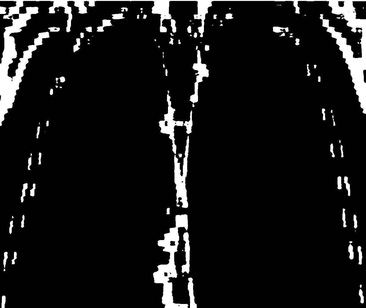
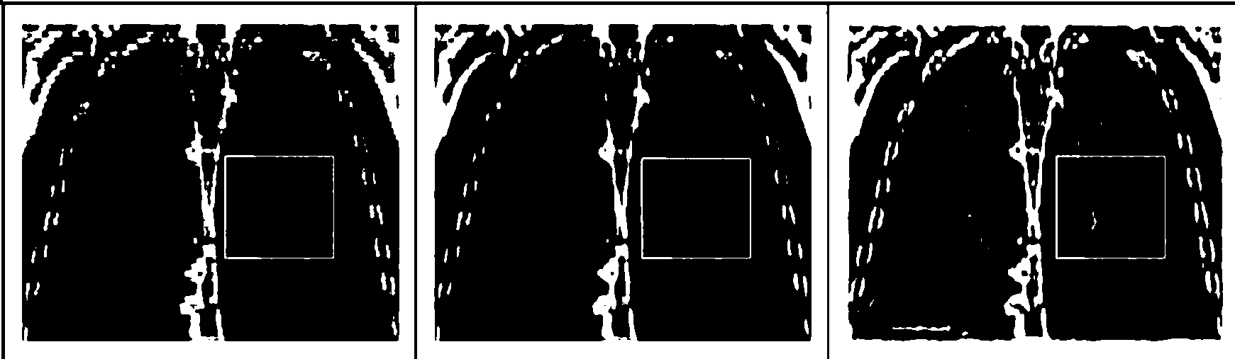
[0103] figure 1 The coronal initial low-resolution image of a certain phase of lung 4D-CT is shown, figure 2 is true figure 1 The image processed by the nearest neighbor interpolation method can be seen from the figure that the ...


PUM


Abstract

The invention discloses a method for reconstruction of a super-resolution coronary sagittal plane image of a lung 4D-CT image based on motion estimation. The method for reconstruction of the super-resolution coronary sagittal plane image of the lung 4D-CT image based on the motion estimation comprises the sequential steps of (1) reading data of the lung 4D-CT image which is formed by a plurality of lung 3D images, wherein the phase positions of the lung 3D images are different; (2) extracting coronary sagittal plane images, corresponding to the same position of the lung, from all the phase positions according to the data of the lung 4D-CT image; (3) estimating motion vector fields between the lung coronary sagittal plane images with different frames based on the full search block matching algorithm; (4) reconstructing the super-resolution lung 4D-CT coronary sagittal plane image by means of the iteration back projection method and based on the motion vector fields obtained in the step (3). According to the method for reconstruction of the super-resolution coronary sagittal plane image of the lung 4D-CT image, the resolution ratio of the reconstructed super-resolution lung 4D-CT coronary sagittal plane image obtained with the method is improved obviously, the brightness and definition of blood vessels and peripheral tissue in the lung parenchyma are improved obviously in a partial enlarged image, the limitation of low resolution caused by the collection time and radiological dose is eliminated, and accurate radiotherapy of lung cancer can be effectively guided.

Description

technical field [0001] The invention relates to the technical field of medical image processing, in particular to a super-resolution coronal sagittal plane image reconstruction method based on motion estimation lung 4D-CT images. Background technique [0002] Lung 4D-CT images can provide comprehensive high-precision radiotherapy respiratory motion characterization. In the lung 4D-CT image data, since there are images of multiple phases, usually 10-20, each phase image is helpful to obtain lung respiratory motion information, which is the key to precise positioning of radiation therapy targets, so lung 4D -CT technology is playing an increasingly important role in precise radiation therapy for lung tumors. [0003] The acquisition of lung 4D-CT data is usually obtained by sorting multiple free-breathing 3D-CT data segments according to the bed location and lung volume. However, dense sampling along the longitudinal direction (commonly named the Z-axis direction) is often i...

Claims


Application Information

Patent Timeline
IPC IPC(8): G06T11/00G06T7/20
Inventor 张煜肖珊
Owner SOUTHERN MEDICAL UNIVERSITY
Why Eureka
Try Eureka