Cytokine-producing cell sheet and method for using same
A cytokine and cell sheet technology, applied in biochemical equipment and methods, animal cells, vascular endothelial cells, etc., can solve the problems of cell necrosis, outflow, and inability to carry out effective transplantation, and achieve high production capacity and cardiac function recovery. high-capacity effect
- Summary
- Abstract
- Description
- Claims
- Application Information
AI Technical Summary
Problems solved by technology
Method used
Image
Examples
Embodiment 1
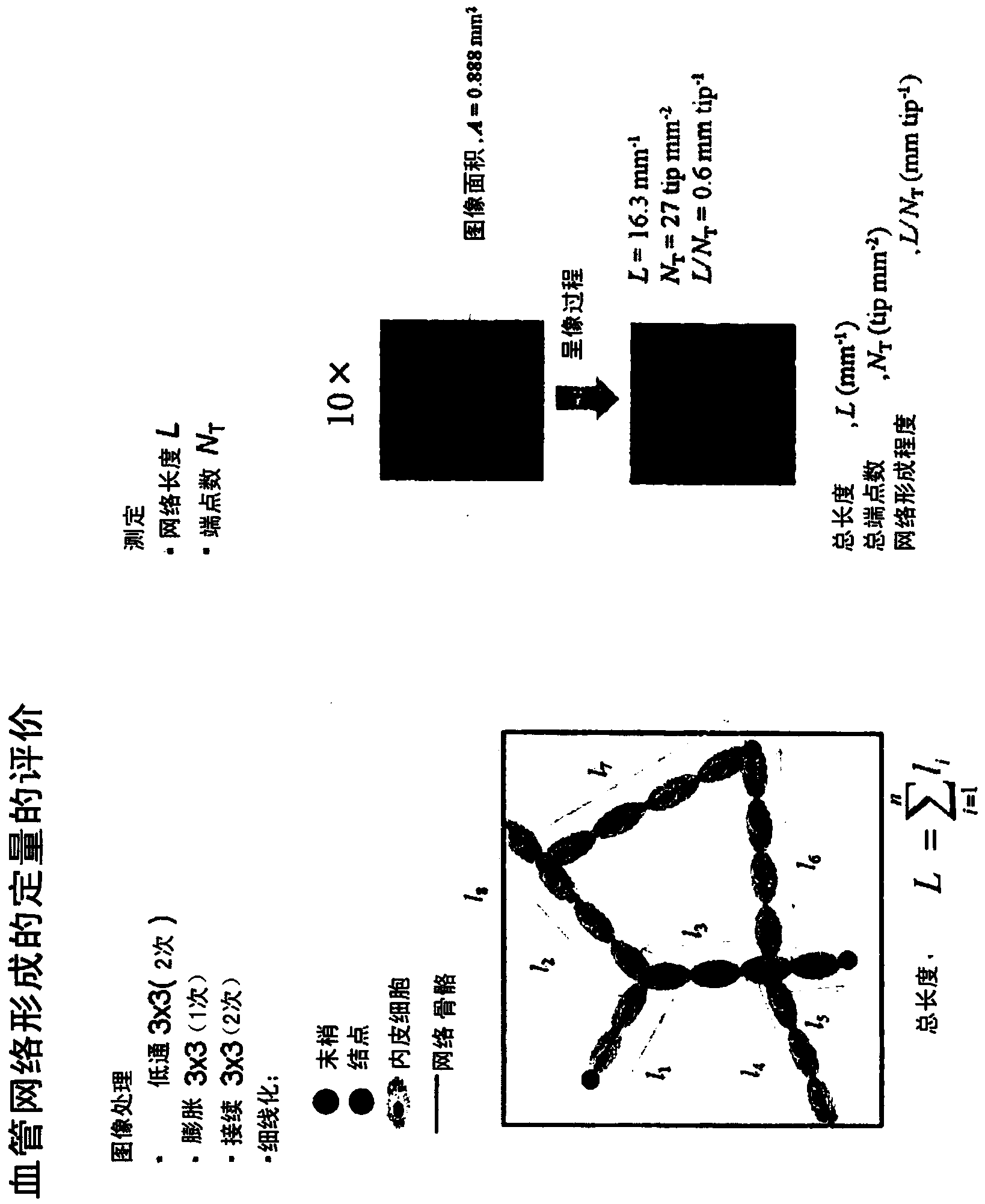
[0075] Construction of a co-culture system of vascular endothelial cells and myoblast sheets (patent document: International Publication No. 2010 / 101225). figure 1 A method for quantitatively evaluating the formation of a vascular endothelial cell network constructed in a cell sheet is shown. The vascular endothelial cell (green) network constructed in the cell sheet is image-processed to deduce the length (L) of the network and the number of endpoints of the network (N T ). The system is calculated by dividing the length of the network (L) by the number of endpoints (N T ) (L / N T ), enabling comparison of the degree of construction of the vascular endothelial network.
[0076] (Analysis of the behavior of vascular endothelial cells in high-density and low-density myoblast sheets)
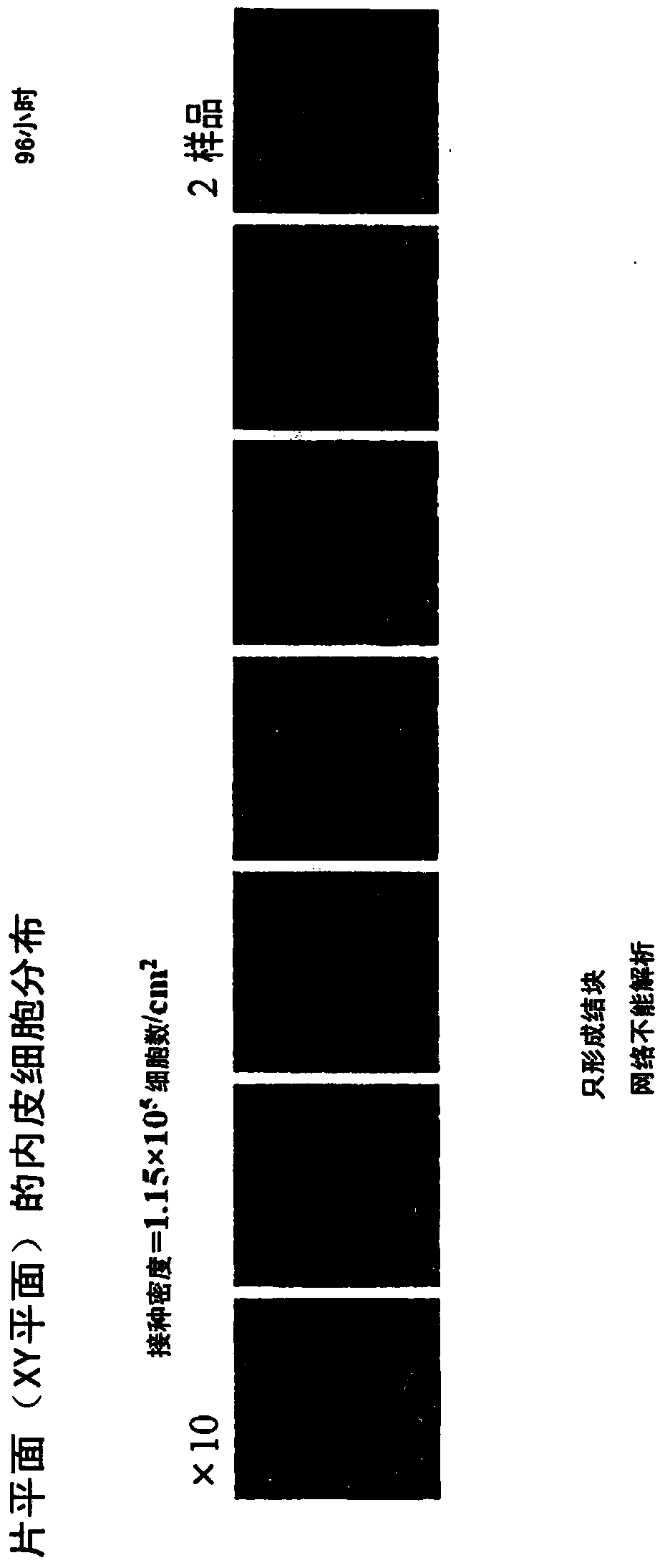
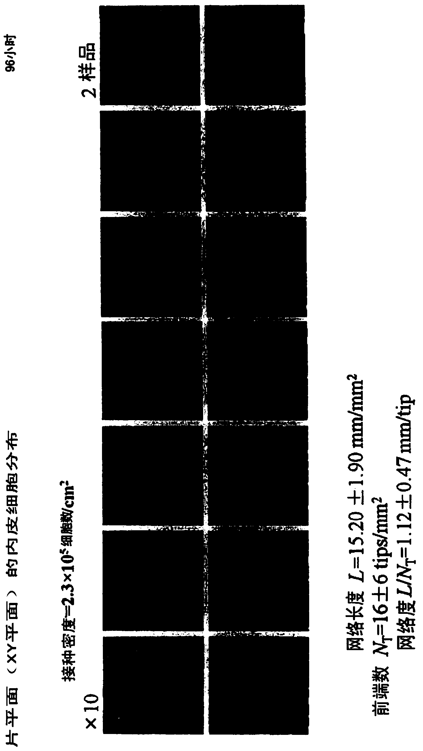
[0077] Using the above-mentioned system, cell sheets with different cell densities were co-cultured with endothelial cells, and the effects of different cell densities on the formation of endo...
Embodiment 2
[0098] (cytokine production capacity of myoblasts dependent on culture state)
[0099] The five-layer cell sheet is a three-dimensional tissue formed by three-dimensionally overlapping single-layer sheets formed by confluence, and it can be said that it is also in a state of dense confluence in three dimensions. Cells temporarily lose proliferative properties due to contact inhibition. In order to understand the cytokine production of sheet-forming cells cultured three-dimensionally in a confluent state, the characteristics of cytokines in a more simple system, that is, normal planar culture, were compared. The culture state can be regarded as a layered structure according to low-density, high-density, sheet, laminated sheet, dense culture of cells, flat and three-dimensional culture. Cytokine production per unit cell was investigated for this hierarchical structure.
[0100] Dilute myoblasts at 1.0×10 4 Cell number / cm 2 , 8.0×10 4 Cell number / cm 2 , 2.3×10 5 Cell numbe...
Embodiment 3
[0124] (for the network formation of endothelial cells in mixed sheets of myoblasts and fibroblasts)
[0125] A myoblast sheet and a myoblast / fibroblast mixed sheet were mounted on endothelial cells for co-culture. It is known that the properties of the cell sheet itself, such as sheet fluidity and cytokine secretion pattern, are adjusted by mixing of fibroblasts, and these are considered to impart adjustments to the surrounding environment to vascular endothelial cells. Therefore, research on the influence of hybrid slices on the formation of the network structure ( Figure 10 ).
[0126] Figure 11 Results of network formation using mixed sheets of skeletal muscle myoblasts and dermal fibroblasts are shown. At 48 hours of co-culture (i.e., 48 hours after the start of the co-culture of skeletal muscle myoblasts and dermal fibroblasts with vascular endothelial cells), 50% of myoblasts were mixed with 100% of myoblasts. % flakes can also form a dense multi-branched homogene...
PUM
| Property | Measurement | Unit |
|---|---|---|
| thickness | aaaaa | aaaaa |
Abstract
Description
Claims
Application Information
 - R&D
- Intellectual Property
- Life Sciences
- Materials
- Tech Scout
- Unparalleled Data Quality
- Higher Quality Content
- 60% Fewer Hallucinations
Browse by: Latest US Patents, China's latest patents, Technical Efficacy Thesaurus, Application Domain, Technology Topic, Popular Technical Reports.
© 2025 PatSnap. All rights reserved.Legal|Privacy policy|Modern Slavery Act Transparency Statement|Sitemap|About US| Contact US: help@patsnap.com


