Positioning ultrathin slice method for pathologic cell infected with virus

A technology for ultra-thin sectioning and diseased cells, which is applied in the preparation of test samples and sampling devices, etc. It can solve the problems of inaccurate positioning, random distribution of target cells, and low detection efficiency, so as to simplify operating procedures and shorten sample preparation time , the effect of reducing randomness

Inactive Publication Date: 2014-02-05
STATION OF VIRUS PREVENTION & CONTROL CHINA DISEASES PREVENTION & CONTROL CENT
View PDF6 Cites 14 Cited by
  • Summary
  • Abstract
  • Description
  • Claims
  • Application Information

AI Technical Summary

Problems solved by technology

[0014] The purpose of the present invention is to solve the problems of random distribution of target cells, inaccurate positioning and low detection efficiency in the existing ultra-thin section technology of cell samples, and to provide a method for positioning ultra-thin section of diseased cells infected with viruses

Method used


Image

  • Positioning ultrathin slice method for pathologic cell infected with virus
  • Positioning ultrathin slice method for pathologic cell infected with virus
  • Positioning ultrathin slice method for pathologic cell infected with virus

Examples


Embodiment 1

[0069] 1. Inoculation of Human Adenovirus

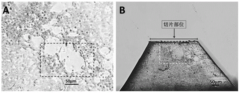
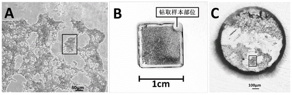
[0070] When subcultured on the Lab-Tek Chamber slide TM When the HEK293 cells in the chamber slide wells of the system (177445) grow to about 80% abundance, inoculate Ad5 on HEK293 cells with cell maintenance medium (DMEM containing 2% FBS) at MOI=0.1. 24-48 hours after inoculating the virus, when the cells develop pathological changes, fix the cells in situ with 2% PFA-2.5% GA (0.1M Caco buffer, pH7.4) at 4°C for 10 minutes; Dehydrate the cells with 90%, 100%, and 100% ethanol for 5 minutes each time; then infiltrate the cells with ethanol:resin ratios of 1:1, 1:4, and 100% resin for 30 minutes each time.

[0071] 2. Resin embedding

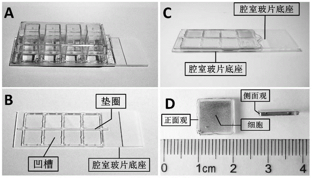
[0072] After infiltrating the cells with the resin, remove the plastic holder of the chamber slide, keep the silicone gasket, add the resin into the square groove formed by the gasket, make the resin plane level with the height of the gasket, and cover the chamber slide base on the gasket , to seal...

Embodiment 2

[0094] 1. Inoculation of influenza virus

[0095] When subcultured on the Lab-Tek Chamber slide TM When MDCK cells grow to about 80% abundance in the chamber slide well of system (177445), inoculate influenza virus Influenza A virus (H1N1 ) on MDCK cells. 24-48 hours after inoculating the virus, when the cells develop pathological changes, fix the cells in situ with 2% PFA-2.5% GA (0.1M Caco buffer, pH7.4) at 4°C for 10 minutes; Dehydrate the cells with 90% and 100% ethanol for 5 minutes each time; then infiltrate the cells with ethanol:resin ratios of 1:1, 1:4 and 100% resin for 30 minutes each time.

[0096] 2. Resin embedding

[0097] After infiltrating the cells with the resin, remove the plastic holder of the chamber slide, keep the silicone gasket, add the resin into the square groove formed by the gasket, make the resin plane flush with the height of the gasket, and take a clean glass slide to cover the gasket , to seal the resin. Using a microwave processor (PELC...


PUM

PropertyMeasurementUnit
thicknessaaaaaaaaaa

Abstract

The invention relates to a positioning ultrathin slice method for a pathologic cell infected with a virus. The method comprises the following steps: (1) with a chamber slide as an embedding die and cells infected with a virus as a model, carrying out in-situ embedding on the cells by using a resin when a lesion occurs in the cells; (2) directly exposing a target cell through trimming or drilling out the target cell and then exposing the target cell through trimming; (3) preparing an ultrathin slice for the target cell; and (4) observing the ultrathin slice of the target cell by using a transmission electron microscope.

Description

technical field [0001] The invention relates to a positioning ultrathin section method for diseased cells infected with viruses, which belongs to the field of ultrathin section sample preparation. Background technique [0002] Transmission electron microscope (TEM) technology is an important means of detecting and studying viruses, and it is also one of the preferred technologies for the diagnosis of unknown pathogens and the identification of pathogens in bioterrorism attacks (References 1, 2). TEM technology has played an important role in the discovery and identification of hepatitis B virus, rotavirus, hantavirus, SARS-Cov and many other viruses (references 3-6). [0003] Negative staining technology and ultrathin section technology are important technologies that make up the biological TEM technology system. Liquid samples are mainly detected by negative staining technology, and solid samples such as tissues and cultured cells are mainly detected by ultrathin section t...

Claims


Application Information

Patent Timeline
Patent Type & Authority Applications(China)
IPC IPC(8): G01N1/06G01N1/28
Inventor 宋敬东屈建国洪涛
Owner STATION OF VIRUS PREVENTION & CONTROL CHINA DISEASES PREVENTION & CONTROL CENT
Why Eureka
Try Eureka