Method for separating circulating tumor cells from blood

A tumor cell and blood technology, applied in the field of biological and medical detection, can solve the problems of low cell purity, cumbersome operation, and difficult long-term storage of chips, and achieve the effect of improving purity, easy operation, and reducing non-specific adsorption

Active Publication Date: 2014-03-19
SHANGHAI JIAO TONG UNIV
View PDF8 Cites 15 Cited by
  • Summary
  • Abstract
  • Description
  • Claims
  • Application Information

AI Technical Summary

Problems solved by technology

However, the capture efficiency of the capture method based on microfluidic chips and antibodies still needs to be further improved, the purity of the captured cells is low, and there is a large amount of non-specific adsorption of white blood cells, and these methods require antibody modification on the chip. It is cumbersome, and the chip loaded with antibodies is not easy to store for a long time

Method used


Image

  • Method for separating circulating tumor cells from blood

Examples


Embodiment 1

[0029] Example 1 Microfluidic chip substrate loaded with polynucleotide

[0030] The polynucleotide with the sequence shown in SEQ ID NO. 1 was dissolved in pure water to prepare a solution with a concentration of 400 μM, and then diluted with dimethyl sulfoxide (DMSO) at a volume ratio of 2:1 to a solution with a final concentration of 267 μM. At the same time, the polydimethylsiloxane PDMS (RTV615, purchased from General Electric Company) microfluidic chip used to infuse polynucleotides was attached to polylysine glass (purchased from Thermo Fisher Scientific Thermo Fisher Scientific company), placed in an oven at 80°C for 2 hours. After the treatment, a constant-flow pump was used to infuse the polynucleotide solution with a concentration of 267μM. After filling, it was placed in a drying oven to dry. After the liquid in the channel was completely dried, the microfluidic chip was removed and the glass was placed in an oven. Treat it at ℃ for 4 hours, then quickly wash out the...

Embodiment 2

[0031] Example 2 Production of "fishbone type" CTC capture chip

[0032] A "fishbone type" PDMS chip with periodic surface unevenness structure (see Wang, ST; Liu, K.; Liu, JA et al, .Angew.Chem.Int.Ed.2011, 50, 3084-3088 for manufacturing method ) Paste on the poly-lysine glass loaded with polynucleotides to ensure that the microchannel of the "fishbone" chip overlaps with the base polynucleotide area, and place the chip in an oven at 80°C for 2 hours. After the treatment is complete After the chip returns to room temperature, fix it with a clamp. The "fishbone" chip has an entrance and an exit. The channel is a continuous S-shaped, with a width of 1mm and an overall length of 200mm. The cross-section of the chip has a periodic uneven structure and is made of two layers of SU8 photoresist. The thickness of the lower photoresist is 70μm, the thickness of the upper photoresist is 35μm, and the fishbone patterns on the upper layer are periodically arranged. The width of the "fishb...

Embodiment 3

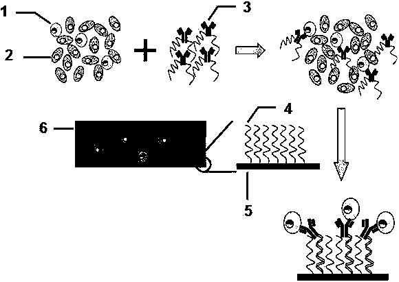
[0033] Example 3 "Fishbone" chip captures rare tumor cells in blood

[0034] First, the "fishbone" chip is perfused with 3% bovine serum albumin (BSA) through a constant flow pump, and the chip is sealed by standing at room temperature for 1 hour. Suspend colon cancer tumor cells HCT116 in the cell culture medium, stain them red with the cell membrane red fluorescent probe DiI (purchased from Biyuntian), and then mix 1000 HCT116 cells with 1 ml of anticoagulated whole blood from healthy people At the same time, add 1 microliter of 1mg / ml epithelial cell adhesion molecule (EpCAM)-labeled single-stranded polynucleotide complex (sequence is shown in SEQ ID NO.2, wherein the 5 end of the sequence, that is, the left end is labeled with amino, will The single-stranded polynucleotide is labeled on the antibody using Solulink's Antibody-Oligonucleotide All-in-One Conjugation Kit), mixed thoroughly for half an hour and then passed through the "fishbone type" with a syringe and a constant ...


PUM


Abstract

The invention discloses a method for separating circulating tumor cells from blood. The method comprises steps of: A. mixing a blood sample with an antibody-labeled single-stranded polynucleotide composite capable of specific binding to a specific antigen on the surfaces of the circulating tumor cells, and incubating; and B. allowing an incubated blood sample to flow through a micro-fluidic chip in which single-stranded polynucleotide is carried at least one surface of microchannels of the chip, separating the circulating tumor cells from the blood through specific binding of the single-stranded polynucleotide to the labeled single-stranded polynucleotide in the antibody-labeled single-stranded polynucleotide composite, and fixing the circulating tumor cells on the surfaces of the chip, thus achieving separation of the circulating tumor cells with the blood sample. The microchannels of the chip have special geometric constructions and can increase the probability that cells in the blood are contacted with the surfaces of the microchannels, thus increasing the capturing efficiency of the circulating tumor cells. Antibody loading is not needed for the chip. The whole capturing process is simple in operation. In addition, the chip is liable to long-term preservation.

Description

Technical field [0001] The invention relates to biological and medical testing, and relates to a method for separating rare cells from a mixed cell population, and in particular to a method for separating circulating tumor cells from a blood sample. Background technique [0002] Rare cells refer to cells that are rare in biological samples but have important biological functions or clinical detection significance, such as circulating tumor cells in human peripheral blood, circulating endothelial cells, cells infected with viruses, and tumors in solid tumors Stem cells etc. Because the number of rare cells is extremely scarce, their detection is like finding a needle in a haystack, and further analysis is even more difficult. Therefore, there is an urgent need to develop methods and tools that can efficiently and quickly separate rare cells from complex biological samples. [0003] Circulating tumor cells (CTC) in human peripheral blood are a representative type of rare cells, whic...

Claims


Application Information

Patent Timeline
Patent Type & Authority Applications(China)
IPC IPC(8): C12N5/09
Inventor 施奇惠邓宇亮张瑜孙帅
Owner SHANGHAI JIAO TONG UNIV
Why Eureka
Try Eureka