MR (magnetic resonance) imaging analysis method for sciatic nerve

An imaging method and technology of sciatic nerve, which is applied in the field of MR imaging analysis of sciatic nerve, can solve the problem that small nerve neuropathy around sciatic nerve cannot be continuously and comprehensively displayed, and achieve the effect of improving feasibility and reliability

Inactive Publication Date: 2014-04-02
THE FIRST AFFILIATED HOSPITAL OF GUANGZHOU MEDICAL UNIV (GUANGZHOU RESPIRATORY CENT)
View PDF2 Cites 5 Cited by
  • Summary
  • Abstract
  • Description
  • Claims
  • Application Information

AI Technical Summary

Problems solved by technology

[0010] The invention provides an MR imaging analysis method for the sciatic nerve, which can overcome the technical defects that the MR ima...

Method used


Image

  • MR (magnetic resonance) imaging analysis method for sciatic nerve
  • MR (magnetic resonance) imaging analysis method for sciatic nerve

Examples


Embodiment 1

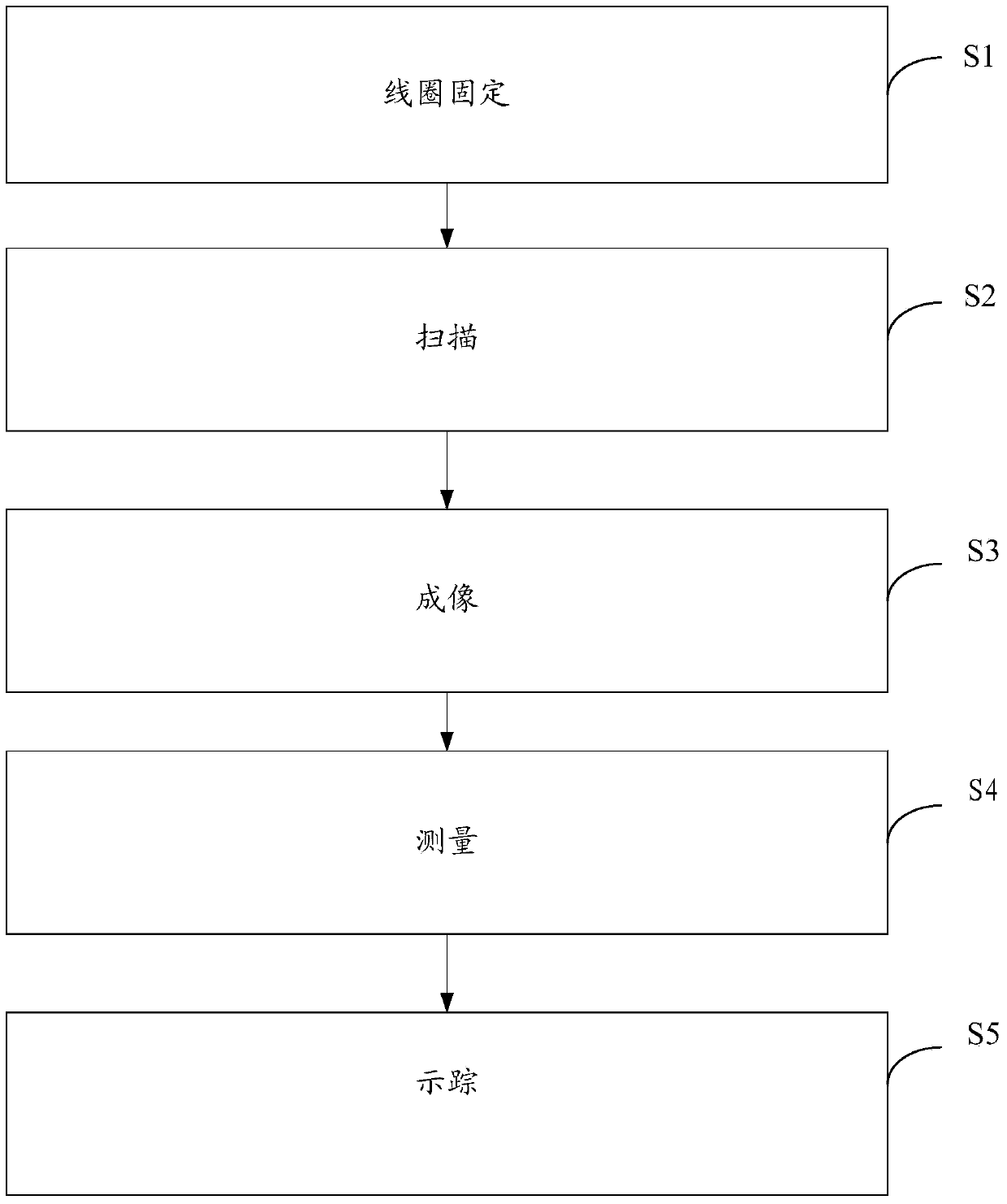
[0053] S1. Coil fixation: Fix the sample to be tested with a phased array radio frequency coil;

[0054] S2. Scanning: scan the sample to be tested fixed by the coil with a dual-gradient nuclear magnetic resonance imaging scanner with a gradient field strength of 66mT / m and a gradient climbing rate of 160T / m / s to obtain scanning signals;

[0055] S3. Imaging: Using sensitive gradient coding and half-Fourier coding technology to collect scanning signals obtained in step S2 for diffusion tensor imaging to obtain data, the sequence is a single-shot spin echo-planar echo sequence, and the diffusion sensitivity coefficient (b value) is 1000s / mm 2 . The repetition time is 4000ms and the echo time is 70ms. The slice thickness / interval is 2mm / 0mm, the scanning field of view is 128x128mm, the scanning matrix is ​​80x80, and the voxel is 1.6mm; the reconstruction matrix is ​​256x256, the reconstruction voxel is 0.5mm, the number of slices is 30, and the scanning time is 9 minutes and ...

experiment example

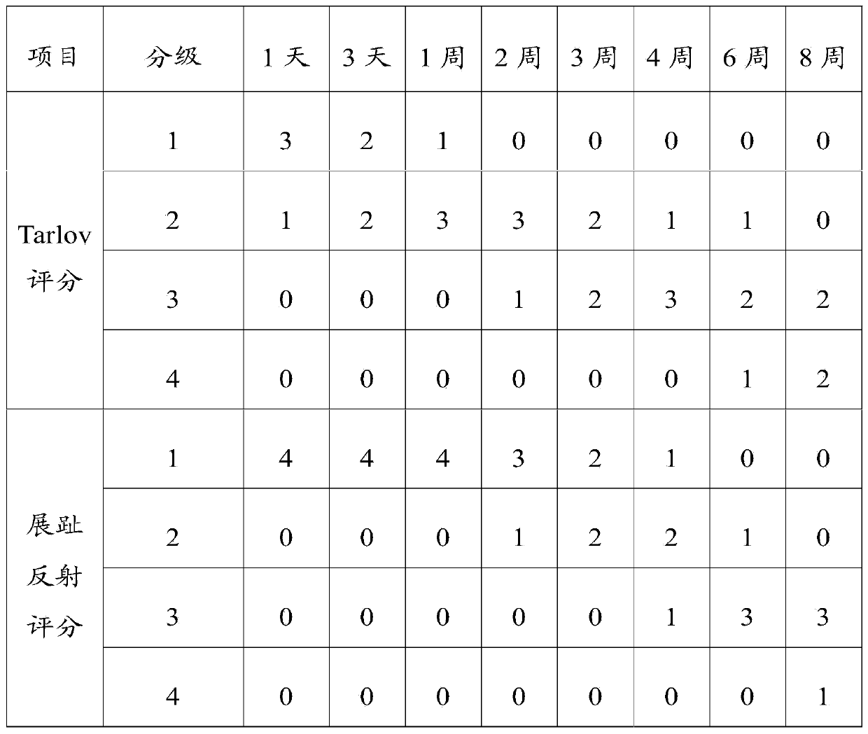
[0059] Select 32 healthy New Zealand white rabbits to conduct experiments according to the method in Example 1, to verify the effectiveness of the method in Example 1.

[0060] Sciatic nerve injury model production.

[0061] Experimental group: 32 healthy New Zealand white rabbits were selected and anesthetized. After successful anesthesia, surgery was performed on the right hind limb of the white rabbit to expose and free the right sciatic nerve. Two dentate vas deferens were used to clamp the two ends of the nerve trunk at the two transverse fingers below the sciatic tubercle. The distance between the two ends was 5 mm. Then use even force to separate the two forceps, and the nerve becomes thinner and longer. When the length of the nerve reaches 10mm, forcefully clamp it for 30 seconds, and then suture the skin of each layer of the right hind limb of the white rabbit.

[0062] Control group: The white rabbits in the experimental group were sham-operated on the left hind lim...


PUM


Abstract

The invention discloses an MR (magnetic resonance) imaging analysis method for sciatic nerve. The MR imaging analysis method includes the steps of fixing with a coil, scanning, imaging, measuring and tracing, and particularly includes fixing a to-be-tested sample by the coil, scanning the to-be-tested sample which is fixed by the coil through a nuclear magnetic resonance imaging scanner to acquire scanning signals, collecting the scanning signals and subjecting the scanning signals to diffusion tensor imaging to obtain an anisotropy index diagram, an apparent diffusion correlation coefficient diagram and an eigenvalue diagram, measuring on the corresponding diagrams to acquire anisotropy index, apparent diffusion correlation coefficient and eigenvalue, performing fiber tracing reconstruction to the original diffusion tensor image to acquire a fiber tracing diagram. The MR imaging analysis method for sciatic nerve can improve sciatic nerve imaging accuracy and reliability.

Description

technical field [0001] The invention relates to the field of nuclear magnetic resonance imaging, in particular to an MR imaging analysis method for sciatic nerve. Background technique [0002] The sciatic nerve is composed of the lumbar nerve and the sacral nerve. It comes from the lumbar 4-5 nerve root and the sacral 1-sacral 3 nerve root. It is the thickest of all nerves, but the sciatic nerve is relatively small and does not run in the same plane. Routine sciatic nerve examination mainly includes: spin echo technique (T1WI weighted image, T2WI weighted image), heavy T2WI weighted imaging (magnetic resonance neuroimaging (MRN)), nuclear magnetic resonance MR background suppression diffusion imaging (DWBIS) and magnetic resonance imaging (MRI) enhanced scan. On conventional imaging sequences, nerve signals are similar to muscle signals. Nerves are displayed as isointensity due to surrounding linear high-intensity fat. Nerves can appear as isointensities on fat-suppressed s...

Claims


Application Information

Patent Timeline
IPC IPC(8): A61B5/055
Inventor 李新春孙翀鹏沈粤春万齐陈镜聪何建勋
Owner THE FIRST AFFILIATED HOSPITAL OF GUANGZHOU MEDICAL UNIV (GUANGZHOU RESPIRATORY CENT)
Why Eureka
Try Eureka