
Function and application of IRF3 (Interferon Regulatory Factor 3) to restenosis after stenting and carotid endarterectomy

A technology for restenosis and vascular stenosis, which can be applied to medical preparations containing active ingredients, peptide/protein ingredients, drug combinations, etc.

Active Publication Date: 2014-05-14
WUHAN UNIV
View PDF0 Cites 2 Cited by
  • Summary
  • Abstract
  • Description
  • Claims
  • Application Information

AI Technical Summary

Problems solved by technology

[0005] Currently, there is no cure for this kind of disease. The main treatment of vascular surgery is vascular reconstruction of occluded segment, including balloon dilation, stent placement, and arterial bypass surgery, etc. However, the incidence of restenosis after vascular reconstruction is high (30 % to 60%), which greatly affected the therapeutic effect, so far restenosis after vascular reconstruction is still a clinical problem

Method used


Image

  • Function and application of IRF3 (Interferon Regulatory Factor 3) to restenosis after stenting and carotid endarterectomy
  • Function and application of IRF3 (Interferon Regulatory Factor 3) to restenosis after stenting and carotid endarterectomy
  • Function and application of IRF3 (Interferon Regulatory Factor 3) to restenosis after stenting and carotid endarterectomy

Examples


Embodiment 1

[0029] Example 1 Mouse Vascular Injury Model (VI) Obtained

[0030] 1. Grouping of experimental animals: WT and IRF3-KO mice aged 8-10 weeks and weighing 24-27g were used and divided into four groups: WT vascular injury group; WT sham operation group; IRF3-KO vascular injury group; IRF3 - KO sham operation group, 60 mice in each group. Twenty mice in each group were sacrificed 7 days, 14 days, and 28 days after the operation, and blood vessels in the injured segment were collected for analysis.

[0031] 2. Operation procedure of mouse vascular injury model:

[0032] 1) Accurately weigh the body weight (g) of the mouse in dynamic mode with an electronic balance, accurately prepare 3% pentobarbital sodium solution with double distilled water, shake gently to dissolve it fully, and use 80mg / kg body weight dose to calculate Accurately extract the corresponding volume of pentobarbital sodium solution with a 1mL syringe, and intraperitoneally inject the anesthetized mouse. After t...

Embodiment 2

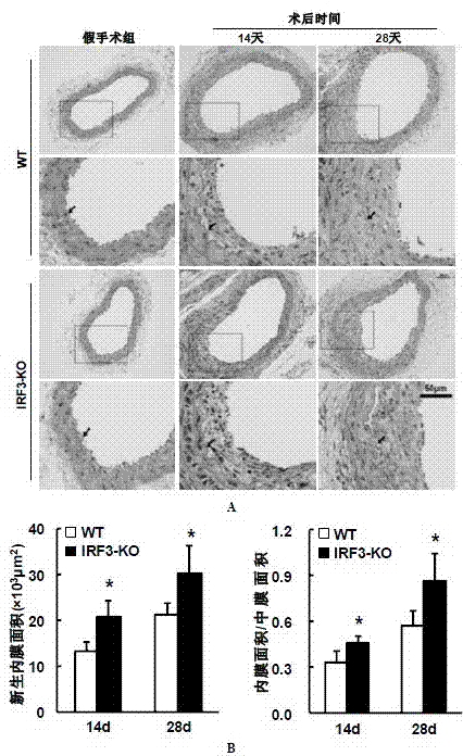
[0037] Example 2 Determination of Intimal Neogenesis in Vascular Injury Model (VI) Mice

[0038] 1. Mice Harvesting

[0039] 1) Anesthetize the mouse, cut the heart and let the blood out.

[0040]2) Cut the carotid artery from the proximal bifurcation of the carotid artery, take 0.5-0.6cm long, and keep the external carotid artery knot.

[0041] 3) Put the carotid artery in PBS, and gently drain the residual blood in the lumen with micro forceps.

[0042] 4) Put the blood vessel into a 1.5mL EP tube filled with 1mL 4% paraformaldehyde for fixation.

[0043] 2. Pathological detection

[0044] 2.1 Preparation of paraffin specimen slices

[0045] Paraffin specimen sections are prepared by professional pathological staff in the laboratory. The main operating procedures include trimming the heart → processing the embedding frame → washing with running water → dehydrating → transparent → dipping in wax → embedding → sectioning (3 μm) → spreading → drying or baking Backup.

[0...

Embodiment 3

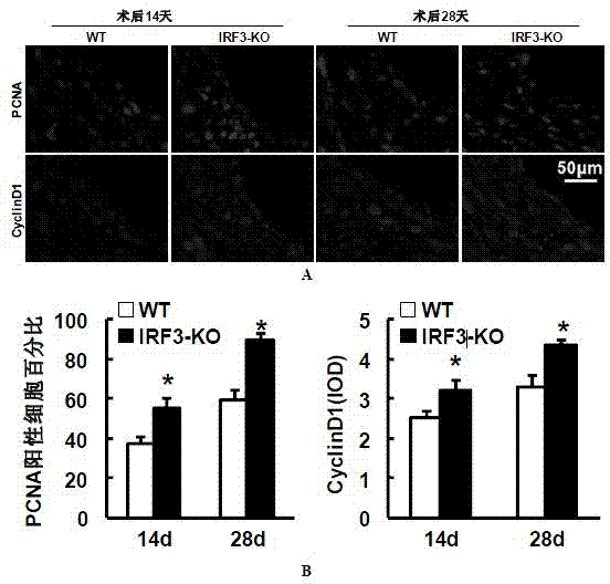
[0053] Example 3 Detection of proliferation level of blood vessel wall cells

[0054] The expressions of proliferating cell nuclear antigen (PCNA) and cell cycle protein (Cyclin D1) were detected by immunofluorescence staining. Required primary antibody information: PCNA (#2586; 1:100; mouse; Cell Signaling Technology), cyclin D1 (#2978; 1:25; rabbit; Cell Signaling Technology); required secondary antibody information: Alexa Fluor 568-conjugated goat anti-rabbit IgG (A11011; Invitrogen, Carlsbad, CA), Alexa Fluor 568-conjugated goat anti-mouse IgG (A11004; Invitrogen, Carlsbad, 150 d, CA).

[0055] The main steps are:

[0056] 1) Baked slices: put the paraffin slices in the oven for more than 30 minutes.

[0057] 2) Dewaxing: Xylene 5min×3.

[0058] 3) Hydration: 100% ethanol 5min×2; 95% ethanol 5min; 70% ethanol 5min; ddH 2 O dipping for 5min×2.

[0059] 4) Citrate tissue antigen repair (high pressure repair): Take a certain amount of pH6.0 citrate antigen repair working...


PUM


Abstract

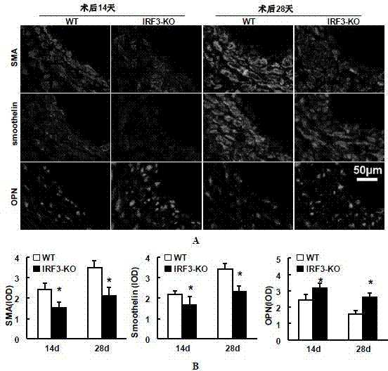
The invention discloses the function and application of an IRF3 (Interferon Regulatory Factor 3) gene to restenosis after stenting and carotid endarterectomy, belonging to the field of function and application of genes. An IRF3 gene knock-out mouse and a wild C57 mouse are taken as experimental objects, and determination of neointima formation, detection of the vascular wall cell proliferation level and detection of smooth muscle cell phenotype of a blood injury model mouse are performed through the blood injury model. As proved by a result, the neointima formation and cell proliferation of the IRF3 gene knock-out mouse are more remarkable than those of the wild mouse. The function of the IRF3 gene to restenosis after stenting and carotid endarterectomy mainly refers to the function of inhibiting restenosis caused by vascular injury of the IRF3 gene, particularly the function of inhibiting restenosis after stenting and carotid endarterectomy. Aiming at the functions of the IRF3, the IRF3 can be used for preparing medicaments for treating restenosis disease, particularly for preparing medicaments for treating restenosis after stenting and carotid endarterectomy.

Description

[0001] technical field [0002] The invention belongs to the field of gene function and application, and relates to the function and application of IRF3 (interferon regulatory factor 3) gene in restenosis after stent and endarterectomy. Background technique [0003] With the change of dietary structure and the aging process of the population, atherosclerotic occlusive lesions are increasing year by year and have become one of the main causes of death in our country. At present, there is no cure for this kind of disease. The treatment methods of vascular surgery include balloon dilation, stent placement and arterial bypass, but restenosis after vascular reconstruction greatly affects the treatment effect. Research on vascular restenosis has been carried out for many years, but so far it is not clear. Studies have shown that in the process of injury formation, the excessive proliferation of neointima and media tissue and the accompanying formation of extracellular matrix are ...

Claims


Application Information

Patent Timeline
IPC IPC(8): A61K38/21A61P9/10
Inventor 李红良张书敏朱丽华张晓东蒋丁胜黄玲
Owner WUHAN UNIV
Learn More