Ultrahigh-sensitivity probe for disease marker or pathogen detection, as well as preparation method and application of ultrahigh-sensitivity probe

A pathogen detection and marker technology, applied in the fields of biomedical engineering and nanomedicine, can solve the problems of expensive reagents and skilled workers, limit the application of early detection of diseases, and limit the wide application, and achieve low cost of reagent consumables, simple detection methods, The effect of high-throughput detection

Inactive Publication Date: 2014-07-16
XIAMEN UNIV
View PDF3 Cites 7 Cited by
  • Summary
  • Abstract
  • Description
  • Claims
  • Application Information

AI Technical Summary

Problems solved by technology

People can use the rapid detection technology of pathogens to grasp the prevalence of infectious diseases, which is of great significance for early and correct clinical decision-making. However, the method widely used in clinical biomarker detection is enzyme-linked immunosorbent assay (hereinafter referred to as ELISA), due to its moderate sensitivity, can only be detected after the level of the biomarker has reached a critical threshold concentr...

Method used


Image

  • Ultrahigh-sensitivity probe for disease marker or pathogen detection, as well as preparation method and application of ultrahigh-sensitivity probe
  • Ultrahigh-sensitivity probe for disease marker or pathogen detection, as well as preparation method and application of ultrahigh-sensitivity probe
  • Ultrahigh-sensitivity probe for disease marker or pathogen detection, as well as preparation method and application of ultrahigh-sensitivity probe

Examples


preparation example

[0040] 1. Preparation example (1): The chemical substances used in the following steps are commercially available.

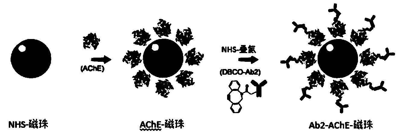
[0041] A preparation of an ultra-high sensitivity probe for disease marker or pathogen detection and a detection method for disease pathogen (taking EV71 as an example), comprising the following steps:

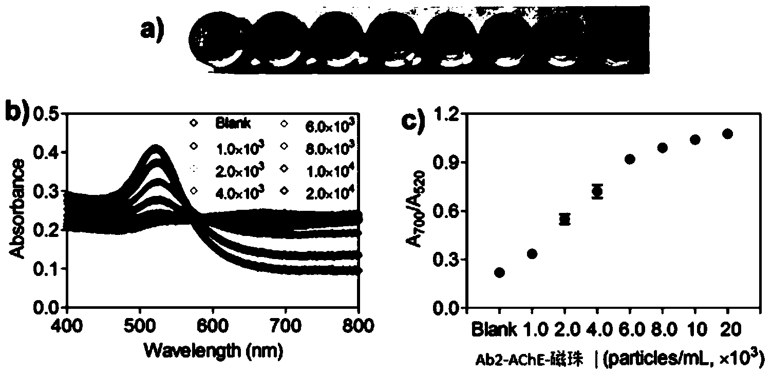
[0042] 1) Take 100 μL of NHS-activated magnetic bead nanoparticles (10 mg / mL) in a small centrifuge tube, and wash it once with hydrochloric acid (1 mM, 4° C.) with the help of a magnetic stand. Afterwards, 100 μL of acetylcholinesterase (1 mg / mL) dissolved in PBS was added, and vortexed for 30 seconds. Afterwards, the mixture was placed in a shaker to react for 2 hours, and the reaction condition was room temperature. For the first 30 minutes, vortex the centrifuge tubes for 15 seconds every 5 minutes. For the remaining 90 minutes, the small centrifuge tube was vortexed for 15 seconds every 15 minutes to obtain magnetic bead nanoparticles bound to acetylcholi...

preparation example (2

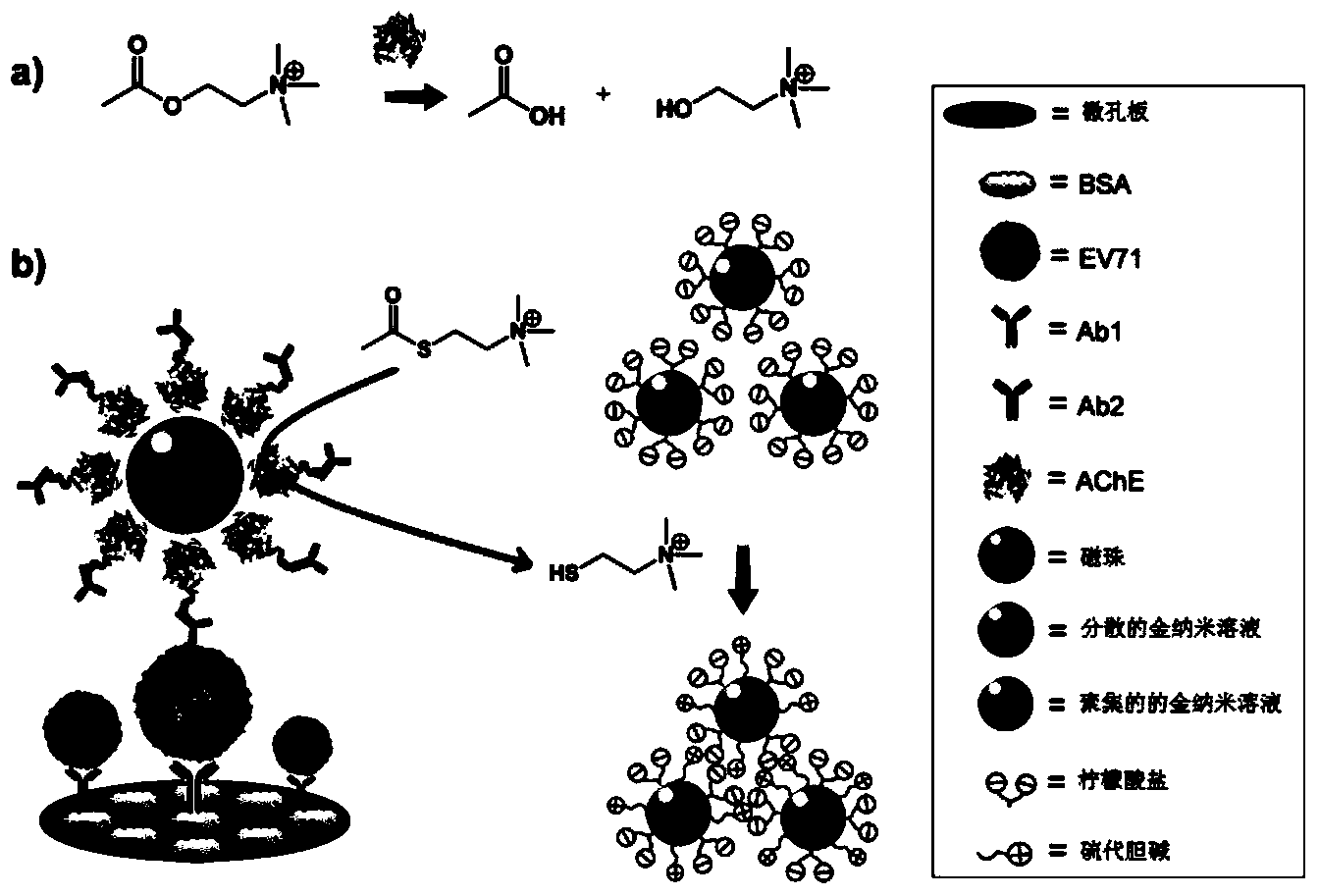
[0051] The preparation of the nanoparticle microsphere probe that does not contain magnetic beads and the detection method used for disease pathogens (taking EV71 as an example) include the following steps:

[0052] 1) Take 100 μL of acetylcholinesterase (1 mg / mL) dissolved in PBS, add 5 μL of NHS-activated azide group (1 mM), and react at room temperature for 30 minutes.

[0053] 2) Take 1 mL of anti-EV71 detection antibody (1 mg / mL), the antibody buffer environment is PBS (pH=7.4), take 33 μL of NHS-activated DBCO (10 mM) and add it to the antibody, and the molar ratio of DBCO to antibody is 50:1. After reacting at room temperature for 30 minutes, 1M Tris-HCl (pH=8.0) was added to terminate the reaction.

[0054] 3) The final concentration of Tris is 50 mM, and the termination reaction lasts for 5 minutes. That is, the antibody solution coupled with DBCO is obtained. Take 10 μL of the antibody solution coupled with DBCO (1 mg / mL) and add it to 1 mL of magnetic bead nanopart...


PUM


Abstract

The invention discloses an ultrahigh-sensitivity probe for disease marker or pathogen detection, as well as a preparation method and application of the ultrahigh-sensitivity probe. The light absorption value of the probe changes based on the collection of a plurality of gold nanoparticles. The probe mainly comprises two major parts, namely bio-functionalized magnetic bead nano microspheres and a gold nanoparticle composite solution. The probe is characterized by containing a detection antibody (Ab2) or a nucleic acid aptamer, biological enzyme active substance surface modified magnetic bead nanoparticles, a substrate which can be catalyzed by enzymes and gold nanoparticles. The ultrahigh-sensitivity probe can be efficiently distinguished from a disease biomarker, and biological enzyme active substances adhered to the surfaces of the magnetic bead nano microspheres can degrade the substrate which can be catalyzed by the biological enzyme active substances into a product with positive charges to collect the gold nanoparticles, so that the light absorption value of the gold nanoparticle solution is changed. According to the method, the detection sensitivity of the disease biomarker is greatly improved; in addition, the method has the advantages of simplify in equipment, simplicity in operation and batch detection capability.

Description

technical field [0001] The invention relates to an ultra-high-sensitivity probe for detection of disease markers or pathogens. Its preparation method and its nanomaterials are used for the detection of disease biomarkers such as tumors or other disease pathogens, and belong to biomedical engineering and nanomedicine. field. Background technique [0002] In terms of in vitro diagnosis of diseases, the detection technology with high specificity, high sensitivity and high throughput characteristics is the goal that people have been pursuing. The establishment of the new rapid detection method simplifies the operation process, reduces the detection cost, and improves the timeliness and accuracy of detection. People can use the rapid detection technology of pathogens to grasp the prevalence of infectious diseases, which is of great significance for early and correct clinical decision-making. However, the method widely used in clinical biomarker detection is enzyme-linked immunos...

Claims


Application Information

Patent Timeline
IPC IPC(8): G01N21/78G01N21/31
Inventor 刘刚陈小元刘定斌王占通杨彩霞
Owner XIAMEN UNIV
Why Eureka
Try Eureka