Application of miRNA to treat myocardial fibrosis diseases
A technology for myocardial fibrosis, disease, applied in the field of biomedical specialty
- Summary
- Abstract
- Description
- Claims
- Application Information
AI Technical Summary
Problems solved by technology
Method used
Image

Examples
Embodiment 1
[0053] Neonatal rat cardiomyocyte culture and cardiomyocyte total RNA and total protein extraction
[0054] l. Primary culture of neonatal rat cardiomyocytes
[0055] 1.1 Take the heart of a newborn rat 1-3 days old and place it in pre-cooled Hanks balanced salt solution (HBSS, Invitrogen).
[0056] 1.2 Separate the myocardial tissue at about 1 / 3 of the lower end of the ventricle, and cut it into small pieces by surgery.
[0057] 1.3 Put a small piece of myocardial tissue into 5 mL of digestion solution containing 75 U / mL collagenase II (Worthington), incubate at 37°C for 20 minutes, collect the digestion solution and add fresh digestion solution to continue digestion. This digestion process was repeated 6 times.
[0058] 1.4 After the cells were centrifuged at 283 g for 5 minutes, they were resuspended in DMEM-F12 cell culture medium (1:1, Invitrogen) containing 10% fetal bovine serum and 1% antibiotics.
[0059] 1.5 Cultivate the cells on the cell culture dish for 1 hour,...
Embodiment 2
[0078] Cardiomyocytes transfected with miR-19b precursor (pre-mir-19b) and stimulated with angiotensin Ang II to induce cardiomyocyte fibrosis
[0079] 1. Extraction of total RNA and total protein after pre-mir-19b transfected cardiomyocytes
[0080] 1.1 Cells were seeded in a 24-well culture plate, the number of cells per well was 4×10 4 .
[0081] 1.2 Mix 1 ul of transfection reagent siPORT NeoFX (AM4510, Ambion) and 25 ul of OPTI-MEM I culture solution (Invitrogen) and incubate at room temperature for 10 minutes.
[0082] 1.3 Dilute pre-mir-19b (AM17100, Ambion) with OPTI-MEM I culture medium (Invitrogen), mix the two and incubate at room temperature for 10 minutes. Using a non-functional miRNA sequence pre-miR TM -Negative Control (AM17110, Ambion) treated cells as a control group (Control).
[0083] 1.4 Mix the diluted pre-mir-19b and transfection reagent, and incubate at room temperature for 10 minutes to form a transfection complex.
[0084] 1.5 Add the culture sol...
Embodiment 3
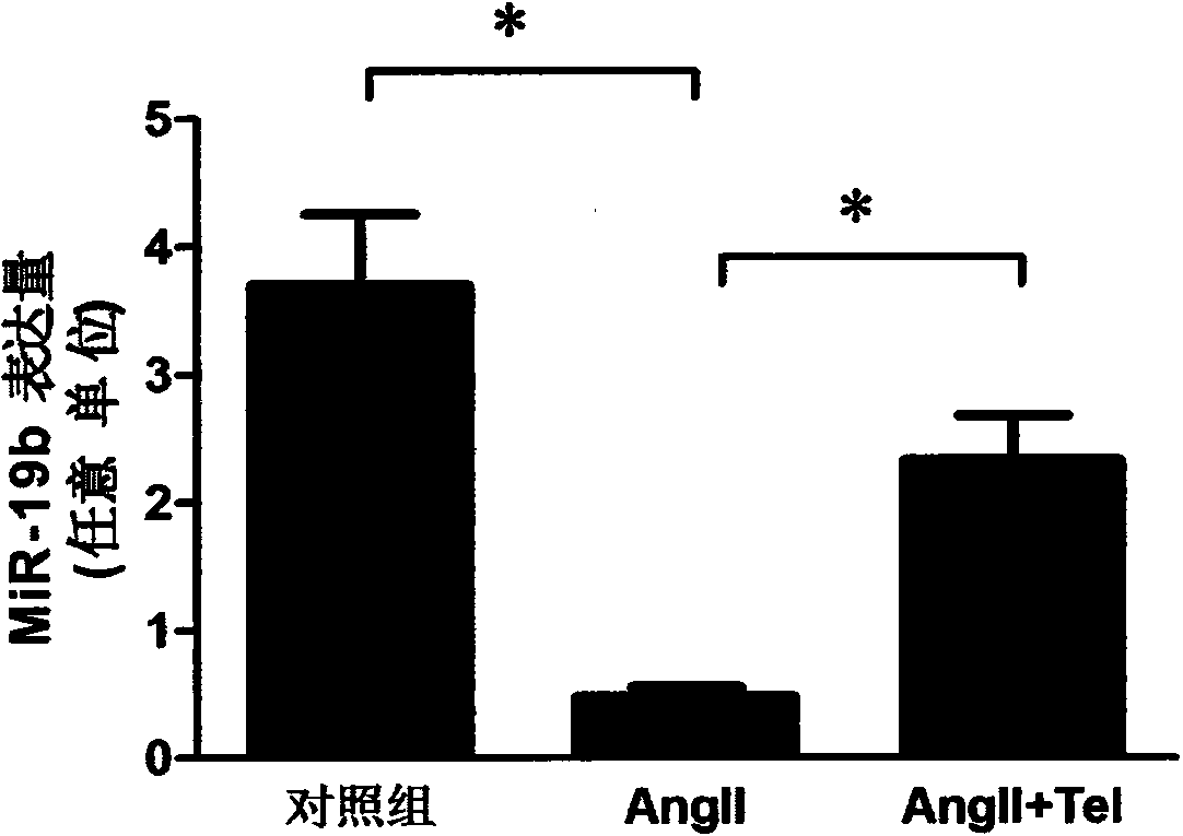
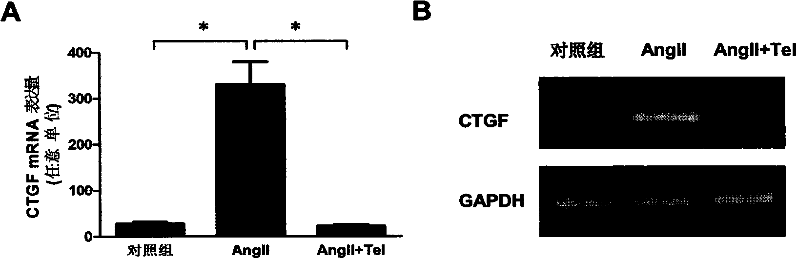
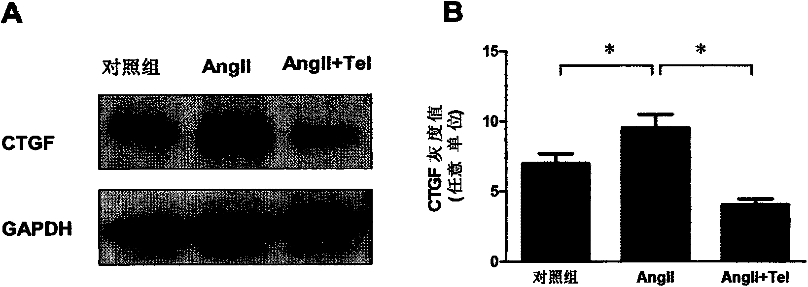
[0093] Analysis of the expression levels of miRNA-19b and CTGF in cardiomyocytes
[0094] 1. qRT-PCR detection of miRNA-19b and CTGF mRNA expression in cardiomyocytes.
[0095] 1.1 Using the collected cardiomyocyte total RNA as a template, the miR-19b gene was amplified with miR-19b-specific mirVana qRT-PCR primers (Ambion). The real-time TaqMan miRNA analysis and detection kit (ABI) was used to perform quantitative PCR to detect the expression of miR-19b in cells, and the U6snRNA gene was used as the internal reference for detection. See the kit instructions for detailed operating principles and methods.
[0096] 1.2 With the total RNA of cardiomyocytes collected as a template, CTGF mRNA was amplified with CTGF gene-specific PCR primers, real-time quantitative polymerase chain reaction (qRT-PCR) was used to detect intracellular CTGF mRNA expression, and GAPDH gene was used as Check the internal reference. PCR products were visualized and quantified by 1% agarose gel electr...
PUM
 Abstract
Description
Claims
Application Information
 - R&D
- Intellectual Property
- Life Sciences
- Materials
- Tech Scout
- Unparalleled Data Quality
- Higher Quality Content
- 60% Fewer Hallucinations
Browse by: Latest US Patents, China's latest patents, Technical Efficacy Thesaurus, Application Domain, Technology Topic, Popular Technical Reports.
© 2025 PatSnap. All rights reserved.Legal|Privacy policy|Modern Slavery Act Transparency Statement|Sitemap|About US| Contact US: help@patsnap.com