Nucleic acid for coding chimeric antigen receptor protein and T lymphocyte for expression of chimeric antigen receptor protein

A technology of chimeric antigen receptors and nucleic acids, applied in the direction of receptors/cell surface antigens/cell surface determinants, genetically modified cells, cells modified by introducing foreign genetic material, etc., can solve tissue damage, off-target cytotoxicity , reducing the activation threshold of effector cells, etc.

Inactive Publication Date: 2014-10-08
CRAGE MEDICAL CO LTD
View PDF9 Cites 28 Cited by
  • Summary
  • Abstract
  • Description
  • Claims
  • Application Information

AI Technical Summary

Problems solved by technology

In addition, excessive co-stimulatory signals in CAR will reduce the threshold required for effector cell activation, so that genetically modified T cells may also be activated under low-level or no antigen-triggered conditi

Method used


Image

  • Nucleic acid for coding chimeric antigen receptor protein and T lymphocyte for expression of chimeric antigen receptor protein
  • Nucleic acid for coding chimeric antigen receptor protein and T lymphocyte for expression of chimeric antigen receptor protein
  • Nucleic acid for coding chimeric antigen receptor protein and T lymphocyte for expression of chimeric antigen receptor protein

Examples


Embodiment 1

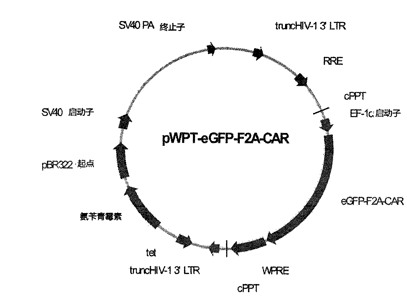
[0035] Example 1. Construction of a lentiviral plasmid expressing the chimeric antigen receptor of the present invention

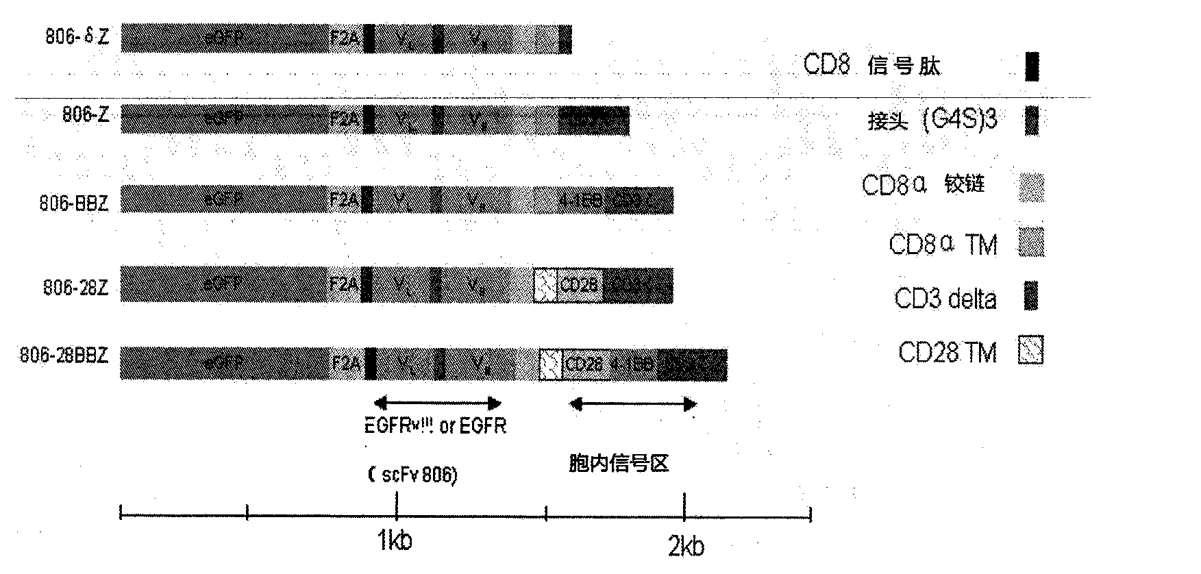
[0036] Table 1 below explains the linkage sequence of the various parts of the exemplary chimeric antigen receptor of the present invention, which linkage can also be found in figure 2 shown in .

[0037] Table 1

[0038] chimeric antigen receptor

Extracellular binding region-transmembrane region-intracellular signal region 1-intracellular signal region 2etc

describe

806-δZ

scFv(EGFR)-CD8-CD3δzeta

negative control

806-Z

scFv(EGFR)-CD8-CD3zeta

first generation

806-BBZ

scFv(EGFR)-CD8-CD137-CD3zeta

second generation

806-28Z

scFv(EGFR)-CD28-CD28-CD3zeta

second generation

806-28BBZ

scFv(EGFR)-CD28-CD28-CD137-CD3zeta

Third Generation

[0039] 1. Amplification of nucleic acid fragments

[0040] (1) Amplification of scFv (EGFR) sequence

[0041] The amplificati...

Embodiment 2

[0084] Example 2. Recombinant lentivirus infection of CD8 + T lymphocytes

[0085] Human peripheral blood mononuclear cells (provided by Shanghai Blood Center) were obtained from the peripheral blood of healthy people by density gradient centrifugation, and peripheral blood mononuclear cells were passed through CD8 + T lymphocyte magnetic beads (Stem Cell Technologies) negative sorting method to obtain CD8 + T lymphocytes, sorted CD8 + T lymphocytes were detected by flow cytometry CD8 + Purity of T lymphocytes to CD8 + The positive rate of T lymphocytes is ≥95% and it is advisable to proceed to the next step. Take about 1 x 10 6 / mL density was added to Quantum007 lymphocyte culture medium (PAA company) for culture, and the magnetic beads (Invitrogen company) coated with anti-CD3 and CD28 antibodies and the final concentration of 100U / mL Recombinant human IL-2 (Shanghai Huaxin Biotech Co., Ltd.) stimulated culture for 24 hours. Then CD8 was infected with the above recom...

Embodiment 3

[0090] Example 3. Detection of EGFR287-302 epitope exposure in epithelial-derived tumor cell lines

[0091] Exposure of the EGFR287-302 epitope on the surface of several tumor cells of epithelial origin was examined using flow cytometry by a fluorescence-activated cell sorter (FACSCalibur, from Becton Dickinson). Materials used include:

[0092] (1) The monoclonal antibody CH12 that recognizes this site constructed by our laboratory (see Chinese patent CN101602808B for the construction method, Examples 1-4) was used as the primary antibody (final concentration 20 μg / ml, 100 μL / sample),

[0093] (2) FITC-labeled goat anti-human IgG is the secondary antibody (AOGMA Company),

[0094] The specific detection method of epitope exposure is as follows:

[0095] 1. Inoculate each tumor cell in the logarithmic growth phase as listed in Table 3 into a 6 cm plate at a cell density of about 90%, and culture overnight in a 37° C. incubator.

[0096] 2. Use 10mM EDTA to digest the cells,...


PUM


Abstract

The invention discloses a nucleic acid for coding a chimeric antigen receptor protein expressed on the surface of the human T lymphocyte. The chimeric antigen receptor protein comprises an extracellular binding domain, a transmembrane domain and an intracellular signal domain, and the extracellular binding domain, the transmembrane domain and the intracellular signal domain are orderly connected. The extracellular binding domain comprises a single-chain antibody scFv (epidermal growth factor receptor EGFR) for specific recognition of 287th-302th amino acid epitopes of EGFR.

Description

technical field [0001] The present invention relates to the field of tumor cell therapy, more specifically, to the field of transgenic T lymphocyte therapy for epithelial-derived tumors expressing EGFRvIII or highly expressing EGFR. Background technique [0002] Epidermal Growth Factor Receptor (EGFR) is a transmembrane glycoprotein with a molecular weight of 170KD. It is the expression product of the proto-oncogene C-erbB-1 (HER-1), and is widely distributed in various tissues of the human body. On the cell membrane [Alan Wells. Molecules in focus EGF receptor. Int J Biochem Cell Biol, 1999, 31: 637-643.]. It is overexpressed and (or ) mutations are closely related to tumor development, malignant transformation, metastasis and prognosis [Jose B. Why the epidermal growth factor receptor? The rational for cancer therapy [J]. Oncologist, 2002, 7(4): 2-8.]. Therefore, EGFR is an important target for tumor therapy. In addition, studies have shown that the EGFR287-302 epitope...

Claims


Application Information

Patent Timeline
IPC IPC(8): C12N15/62C12N15/867C12N7/01C12N5/10C07K19/00
CPCC07K14/7051C07K16/2863C07K2319/74C12N5/0636C12N15/86C12N2510/00C12N2740/15043
Inventor 李宗海高慧萍蒋华石必枝顾健人
Owner CRAGE MEDICAL CO LTD
Why Eureka
Try Eureka