
Microfluidic chip and cell screening method for screening specific cells

A microfluidic chip and specific technology, applied in the field of cell analysis, can solve the problems of difficult CTCs cell screening, difficult to achieve early diagnosis of cancer metastasis, affecting the efficiency of cell screening, cell activity, etc.

Active Publication Date: 2014-12-10
广州康臣药业有限公司
View PDF5 Cites 52 Cited by
  • Summary
  • Abstract
  • Description
  • Claims
  • Application Information

AI Technical Summary

Problems solved by technology

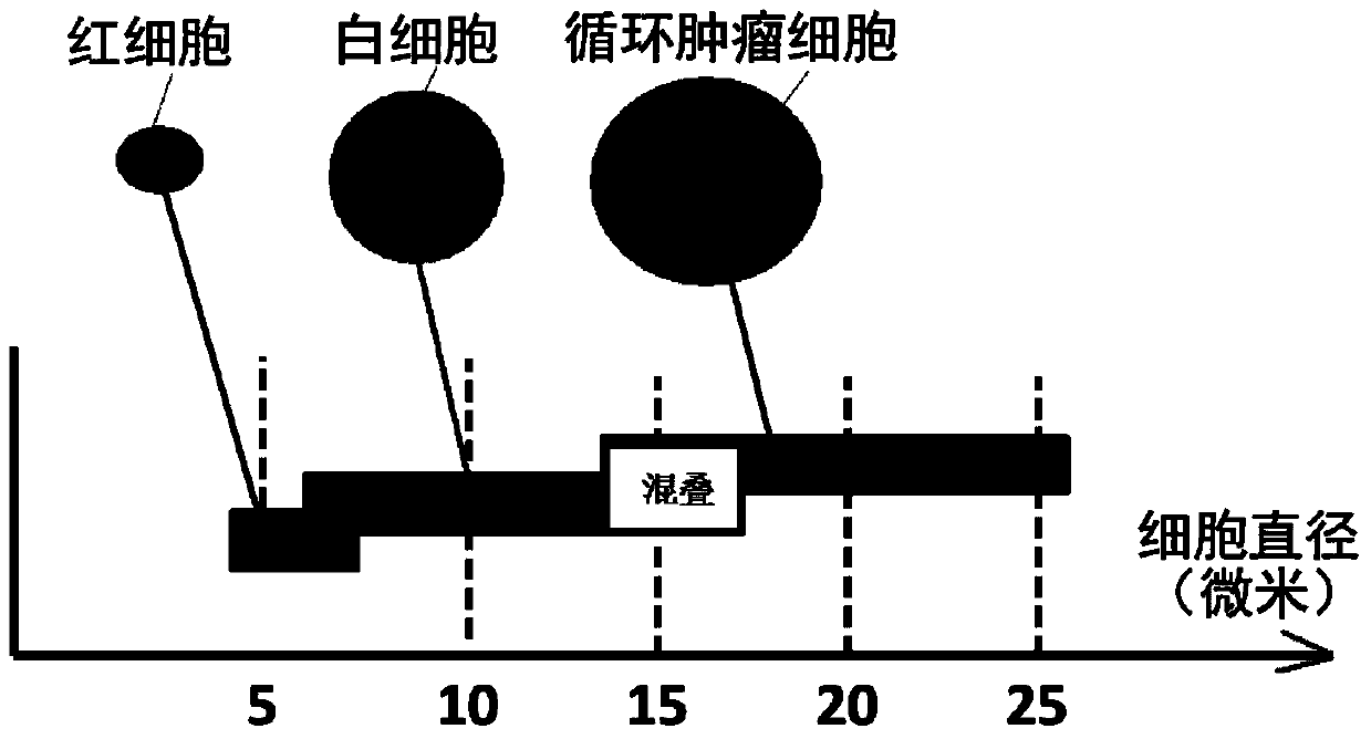
However, the acoustic method lacks specificity, and the particle size of CTCs is aliased with the cell particle size in blood (such as figure 1 shown), it is difficult to screen CTCs cells
[0009] In summary, although there are many methods to screen CTCs, each method has certain shortcomings and deficiencies, which directly affect the efficiency of cell screening and cell activity, making it difficult to achieve early diagnosis of cancer metastasis.

Method used


Image

  • Microfluidic chip and cell screening method for screening specific cells
  • Microfluidic chip and cell screening method for screening specific cells
  • Microfluidic chip and cell screening method for screening specific cells

Examples


Embodiment 1

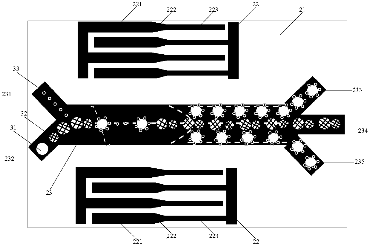
[0027] Such as figure 2As shown, this embodiment provides a microfluidic chip for screening specific cells, including a substrate 21, a cavity 23 attached to the substrate 21, and an acoustic wave excitation source.

[0028] In a specific implementation, in order to obtain a larger electromechanical coupling coefficient, the substrate is a 128°YX double-sided polished lithium niobate crystal.

[0029] In yet another specific implementation, the cavity is made of polydimethylsiloxane (PDMS). The lumen 23 includes a middle channel, inlets 231 , 232 at one end of the middle channel, and outlets 233 , 234 , 235 at the other end of the middle channel. Among them, the inlet 231 is used for injection of targeted microbubbles 33, the inlet 232 is used for injection of blood containing specific cells 31 and blood cells 32 (including red blood cells, white blood cells and platelets), and the outlets 233 and 235 are used for the isolated specific cells. The sex cells and the microbubb...

Embodiment 2

[0041] This example provides a method for screening specific cells, using the microfluidic chip provided in Example 1 as the experimental platform, enriching CTCs under the action of sound waves, and relying on the adhesion of targeted microbubbles to CTCs , enhance the acoustic impedance difference of CTCs, realize the screening of cells by adjusting the amplitude and frequency of the acoustic wave, and the biological activity of cells will not be affected, and propose a new way for the screening of CTCs.

[0042] The process of the cell screening method in this embodiment includes the following steps S1-S4:

[0043] Step S1, preparing targeted microvesicles. Microbubbles, commonly known as ultrasound contrast agents, are encapsulated microbubbles containing inert gases. The primary function of microbubbles is to enhance the contrast of ultrasound images and improve image quality. Recent studies have shown that the surface of microbubbles can be chemically modified, and the ...


PUM


Abstract

The invention discloses a microfluidic chip and a cell screening method for screening specific cells. The chip comprises a substrate, a cavity and an acoustic excitation source, wherein the cavity comprises a middle channel, an inlet and an outlet; the acoustic excitation source is positioned on the substrate and at two sides of the middle channel and comprises two parts which extend to a direction of the outlet along the middle channel, wherein the corresponding acoustic resonance frequency of the second part close to the outlet is integer multiples of the corresponding acoustic resonance frequency of the first part far away from the outlet. Based on an acoustic streaming effect formed by targeted microbubbles and asymmetric sound fields, the microbubbles are adhered to the surfaces of the specific cells, so that the enriched cells which flow past the first part can still move in a straight line and cannot diffuse due to a laminar flow characteristic of microfluidics, and the specific cells bonded with the targeted microbubbles can shift because the acoustic resonance frequency of the second part is different from that of the first part. Thus, the screening of the specificity of the specific cells in blood is realized.

Description

technical field [0001] The invention relates to the technical field of cell analysis, in particular to a microfluidic chip for screening specific cells and a cell screening method. Background technique [0002] Molecular biology and clinical studies have shown that some cancers diagnosed as early stages actually have distant metastases, that is, tumor micrometastases, which are difficult to detect by conventional imaging, histology or cytology methods. Tumor micrometastasis can form micrometastasis lesions in the tissues and organs of the whole body through the hematogenous and lymphatic pathways, and the development of lymph node metastasis will eventually enter the blood circulation to form circulating tumor cells (Circulating Tumor Cells, CTCs), leading to systemic metastasis of the primary tumor. tendency. The detection of CTCs is helpful for early detection of tumor micrometastasis, monitoring of postoperative recurrence, evaluation of curative effect and prognosis, or...

Claims


Application Information

Patent Timeline
IPC IPC(8): C12M1/00C12M1/42C12N5/09
CPCB01L3/502753B01L2200/0668B01L2300/0877B01L2400/0439C12M47/04C12N5/0693C12N2509/00
Inventor 郑海荣孟龙蔡飞燕牛丽丽李飞肖杨
Owner 广州康臣药业有限公司
Learn More