Osteogenic differentiation of mesenchymal stem cells

A technology of osteogenic differentiation and mesenchymal stem cells, applied in animal cells, vertebrate cells, cell culture active agents, etc., can solve the problem of poor understanding of recipient cell differentiation

Inactive Publication Date: 2015-04-01
BIOMATCELL
View PDF4 Cites 3 Cited by
  • Summary
  • Abstract
  • Description
  • Claims
  • Application Information

AI Technical Summary

Problems solved by technology

[0005] However, little is known about whether and how EVs affect the differentiation of recipient cells

Method used


Image

  • Osteogenic differentiation of mesenchymal stem cells
  • Osteogenic differentiation of mesenchymal stem cells
  • Osteogenic differentiation of mesenchymal stem cells

Examples


example 1

[0060] General description of the process

[0061] Isolation and culture of monocytes: Monocytes were isolated from human blood using magnetic separation and cultured on different biomaterials with or without stimulation (eg LPS). Mesenchymal stem cells are obtained from human bone marrow by gradient separation. After 72 h, cells were harvested and exosomes were isolated from conditioned media.

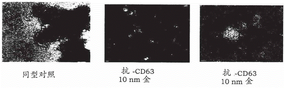
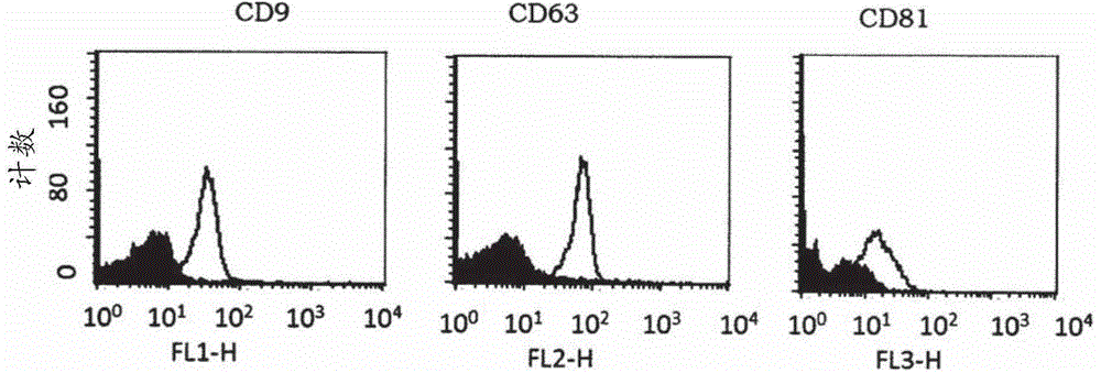
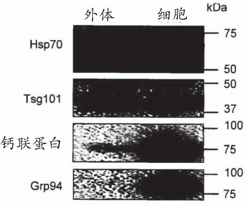
[0062] Exosome Isolation and Detection: Exosomes will be isolated using a method based on repeated centrifugation and filtration steps to remove cellular debris, apoptotic bodies, etc., followed by ultracentrifugation to pellet exosomes. Alternative separation methods can also be used. Exosomes will be detected using a combination of methods including electron microscopy as well as detection of markers commonly found on exosomes (e.g., CD9, CD63, CD81, Tsg101) and a marker that should not be present in exosomes (calnexin).

[0063] mRNA and microRNA content of exosomes: Microarray...

example 2

[0074] Human primary LPS-stimulated monocytes. After 3 days in culture, exosomes were isolated and RNA was extracted. Bioanalyzer analysis of cellular and exosomal total RNA and small RNA. The electropherogram shows ( Figure 4 ) (a) Nucleotide size distribution (nt) and fluorescence intensity (FU) of total RNA in exosomes and cells (b) and small RNAs in exosomes (c) and cells (d).


PUM


Abstract

The present invention relates to a method for inducing and/or promoting osteogenic differentiation using extracellular vesicles and the use thereof.

Description

technical field [0001] The present invention relates to a method of inducing or promoting cell differentiation, extracellular vesicles for use in inducing or promoting said differentiation, uses of said vesicles and a therapeutic method using said vesicles. Background technique [0002] The mechanisms of early bone formation at the implant surface or at the site of injury and the factors affecting maintenance of bone-implant contact, stability and function are not fully understood. Increased knowledge of the pathways by which inflammatory and stem cells communicate with progenitor cells on the surface of implants is important for understanding how the next generation of implants should be optimized for clinical use. With more knowledge, in the future we will hopefully be able to produce new and better implants for improved osseointegration or better drugs for bone healing. [0003] The monocyte / macrophage system plays a central role in host defense, wound healing, and immun...

Claims


Application Information

Patent Timeline
Patent Type & Authority Applications(China)
IPC IPC(8): C12N5/0775A61K35/28A61L27/34A61L27/38A61P19/08A61K35/15
CPCC12N5/0635C12N2502/1157A61K35/15A61L31/005A61L31/16A61L24/0005A61L24/0015A61L2300/64A61L2430/02A61P19/00A61P19/02A61P19/08A61P19/10A61P29/00A61K35/32C12N5/0654C12N2501/052C12N2506/1384
Inventor 卡琳·埃克斯特伦彼得·汤姆森尤卡·劳斯马欧玛·欧玛王晓勤
Owner BIOMATCELL
Why Eureka
Try Eureka