Bio-adhesive agent comprising surface-modified hydroxyapatite and use thereof

a technology of hydroxyapatite and bioadhesive agent, which is applied in the preparation of isocyanic acid derivatives, group 5/15 element organic compounds, organic chemistry, etc., can solve the problems of inability to apply sutures to tissue having a structure that cannot be sutured, tissue that makes up cartilage or bone limitations, etc., and achieve excellent adhesive effect and promote bone regeneration

Active Publication Date: 2013-03-28
UNIV IND COOP GRP OF KYUNG HEE UNIV
View PDF2 Cites 1 Cited by
  • Summary
  • Abstract
  • Description
  • Claims
  • Application Information

AI Technical Summary

Benefits of technology

[0017]Differently from the previous bio-adhesive agents, the bio-adhesive agent comprising the surface-modified hydroxyapatite as an active ingredient according to the present invention can achieve an adhesive effect with full integration such that disconnection between tissues to be adhered does not occur.
[0018]In addition, since the active ingredient hydroxyapatite itself is capable of osteogenesis, when the bio-adhesive agent of the present invention is applied between bone and cartilage or bone and tissue, it can promote regeneration of bone locally at the site to which cartilage or tissue is attached. Thus, it is expected that the bio-adhesive agent of the present invention would exhibit an excellent adhesive effect between bone and cartilage or bone and tissue.
[0019]Furthermore, if the surface of a prosthesis made by metal such as titanium is coated with the bio-adhesive agent of the present invention, the surface-modified hydroxyapatite is attached to the surface of the metal prosthesis and this is expected to resolve the problem of integration with natural tissues, which is a drawback in using the previous metal prosthesis.

Problems solved by technology

However, in case the transplanted cartilage tissues do not integrate into the existing tissues, a full therapeutic effect cannot be expected.
It is impossible to apply a suture to a tissue having a structure that cannot be sutured.
For example, a tissue that makes up cartilage or bone has limitations for having a suture applied thereon.
In particular, when a connection between bone and cartilage is necessary as is in, for instance, the articular cartilage transplantation, it is not easy to carry out such a suture method and there is the shortcoming that a suture area has the potential to cause inflammation.
Furthermore, in the case of widely occurring cartilage damage such as degenerative arthritis, it is not easy to find a way to achieve a connection of tissues over a large area.
However, this agent has a major disadvantage due to the fact that the applied adhesive materials formed insulation between the two attached cartilage tissues making it impossible to communicate or exchange the nutrient and / or growth factors and thus two different disconnected cartilage layers are formed bordering the attached surface.
Moreover, the previously developed bio-adhesive agents are not effective in achieving a connection (adhesion) between bone and cartilage in the full-thickness wound.
However, WO 2009 / 073068 teaches only the combination with a second ingredient such as chitosan and does not teach a use of the surface-modified hydroxyapatite alone.

Method used


Image

  • Bio-adhesive agent comprising surface-modified hydroxyapatite and use thereof
  • Bio-adhesive agent comprising surface-modified hydroxyapatite and use thereof
  • Bio-adhesive agent comprising surface-modified hydroxyapatite and use thereof

Examples


example 1

Surface Modification of Hydroxyapatites and Confirmation

[0067]Glycidyloxypropyl trimethoxysilane (GPMS) or aminopropyltriethoxysilane (APS)-terephthaldicarboxaldehyde (TPDA) was used as a silane-based linker compound, and ethylisocyanate (EI) was used as an isocyanate-based linker compound. These linker compounds and hydroxyapatite crystal powders were added to toluene and refluxed at 110° C. for 1 hour so that the linker compounds were attached to the surface of the hydroxyapatites via a silane or isocyanate group. This procedure is shown in FIG. 1. The first one is a synthesis process of HA-GPMS, the second one is a synthesis process of HA-EI, and the third one is a synthesis process of HA-TPDA.

[0068]In order to confirm a coupling between aldehyde groups on the surface of the surface-modified hydroxyapatites and amine groups, HA-TPDA and general hydroxyapatites (bare-HA) were each mixed with an aqueous solution of chitosan. As a result, as shown in FIG. 2, in the case of HA-TPDA, ...

example 2

Confirmation of Presence of Amine Groups on the Surface of Natural Tissues

[0069]In order to achieve reaction with HA-TPDA, amine groups must be present on the surface of tissues to be attached. Whether or not amine groups are present on the surface of the subject tissues to be attached was determined as follows.

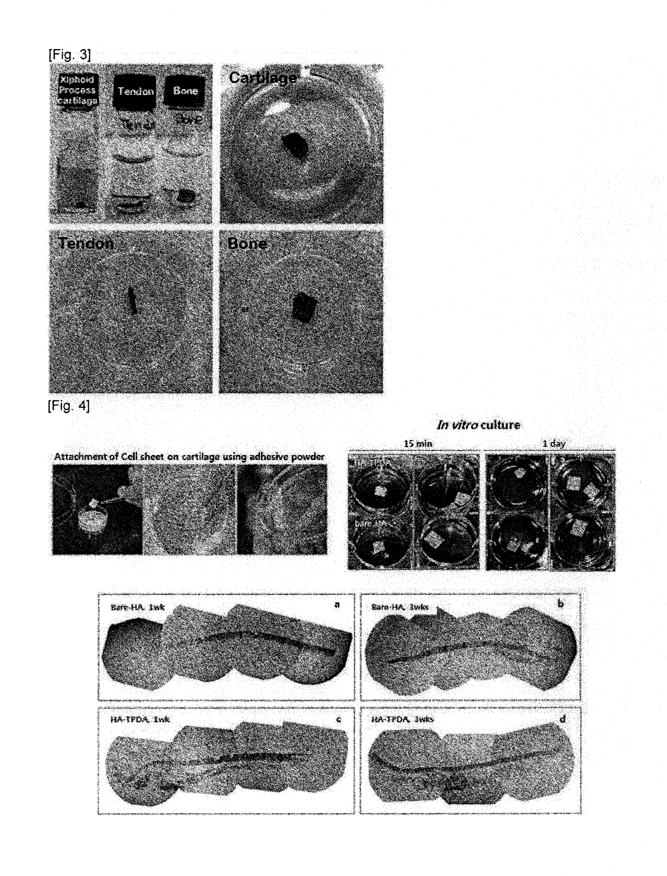
[0070]Cartilage, bone and tendon tissues were carefully isolated from a mouse so that connective tissues were not included. As shown in FIG. 3, the isolated tissues were added to 6% ninhydrin in 98% ethanol, heated at 96° C. for 10 minutes and determined on whether or not the tissues exhibit color. As a result, it was confirmed that the cartilage, bone and tendon tissues have amine groups.

example 3

In vitro Culture Experiment in Order to Confirm the Adhesive Ability of HA-TPDA

[0071]HA-TPDA powders were sprinkled on the cartilage tissues obtained from rabbit ears, and unbound HA-TPDA powders were brushed out. Multiple layers of bone marrow stromal cells (BMSCs) obtained from rats and cultured in the form of a sheet were attached to the tissues. The tissues were cultured in an alpha-MEM medium containing 20% fetal bovine serum, 1% penicillin / streptomycin, 2 mM L-glutamine, 10−8M dexamethasone and 10−4M ascorbic acid for 1 week to 3 weeks, and fixed with 4% paraformaldehyde in PBS. The fixed tissues were embedded in paraffin and cut. The degree of tissue adhered was determined by histological staining, and the results are shown in FIG. 4. As shown in FIG. 4, it was confirmed that the sheet form was remained for a longer time in the HA-TPDA-applied group than in a general hydroxyapatite-applied group.


PUM

PropertyMeasurementUnit
adhesionaaaaaaaaaa
structureaaaaaaaaaa
areaaaaaaaaaaa

Abstract

The present invention relates to a bio-adhesive agent comprising a surface-modified hydroxyapatite and its use. More specifically, the present invention relates to a bio-adhesive agent for the adhesion between bone and bone, bone and tissue, bone and cartilage, or bone and tendon, or for the adhesion of a shield between bones or of an artificial joint, which comprises a surface-modified hydroxyapatite as an active ingredient, wherein the surface-modified hydroxyapatite is characterized in that a certain linker compound is covalently bonded to the surface of the hydroxyapatite; a method for coating the surface of a metal prosthesis using the surface-modified hydroxyapatite; and a metal prosthesis coated with the surface-modified hydroxyapatite obtained by said method.

Description

TECHNICAL FIELD [0001]The present invention relates to a bio-adhesive agent comprising a surface-modified hydroxyapatite and its use. More specifically, the present invention relates to a bio-adhesive agent for the adhesion between bone and bone, bone and tissue, bone and cartilage, or bone and tendon, or for the adhesion of a shield between the bones or of an artificial joint, which comprises a surface-modified hydroxyapatite as an active ingredient, wherein the surface-modified hydroxyapatite is characterized in that a certain linker compound is covalently bonded to the surface of the hydroxyapatite; a method for coating the surface of a metal prosthesis using the surface-modified hydroxyapatite; and a metal prosthesis coated with the surface-modified hydroxyapatite obtained by said method.BACKGROUND ART [0002]Tissue-engineering methods for the repair of damaged tissue include (i) forming a tissue from expanded and cultured cells, and transplanting said tissue, (ii) loading expand...

Claims


Application Information

Patent Timeline
Patent Type & Authority Applications(United States)
IPC IPC(8): A61L24/00
CPCA61L24/02A61L24/00A61L2400/18A61L27/32
Inventor SON, YOUNG SOOKLEE, EUN AHNAM, SEUNG WOO
Owner UNIV IND COOP GRP OF KYUNG HEE UNIV
Why Eureka
Try Eureka