A method for rapid isolation of exosomes

A fast technology for separating cells, applied in animal cells, tumor/cancer cells, vertebrate cells, etc., can solve the problems of high cost and achieve the effects of shortening time, improving the purity of exosomes, and high yield

Active Publication Date: 2017-07-28
FUZHOU HOSPITAL FOR INFECTIOUS DISEASE
View PDF5 Cites 0 Cited by
  • Summary
  • Abstract
  • Description
  • Claims
  • Application Information

AI Technical Summary

Problems solved by technology

This separation method also has higher cost, and the whole separation process takes about 13.5 hours

Method used


Image

  • A method for rapid isolation of exosomes
  • A method for rapid isolation of exosomes

Examples


Embodiment 1

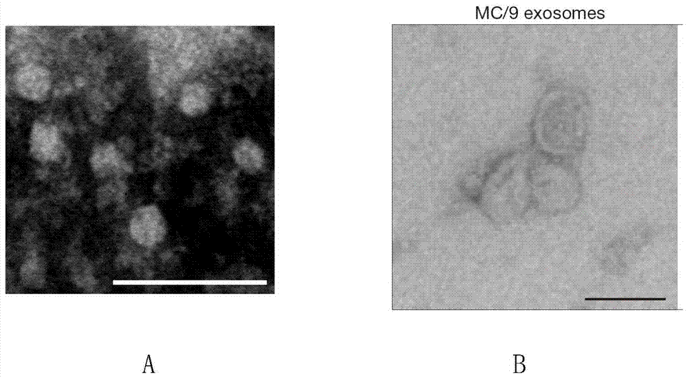
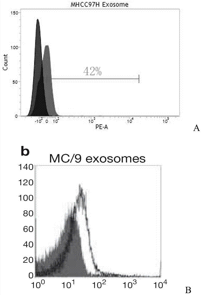
[0027] Example 1: Isolation of extracellular exosomes

[0028] Take human MHCC97H cells as an example (but the method of the present invention is not limited to MHCC97H cells):

[0029] will be 5×10 8 MHCC97H cells (Shanghai Medical College of Fudan University, Fan Jia's research group) were inoculated in T175 (Corning, USA) cell culture flasks containing 60ml of medium, and the medium composition was DMEM (Dulbecco's Modified Eagle Medium, Life technologies, USA) plus 10 % (v / v) of fetal bovine serum (FBS, Life Technologies, USA). At 37°C with 5% CO 2 cultured in an incubator for 48 hours.

[0030] Collect 40 mL of the human liver cancer cell line MHCC97H cell culture supernatant, centrifuge at 400 g at 4°C for 5 min to remove cell debris, add RNase (Sigma, USA) at a final concentration of 1 mg / ml, 6.5% glycerol and 5.5 mM sorbitol, Then use a vacuum centrifugal concentrator (CentriVap, USA) to concentrate by centrifugation at 16° C. at 1500 g for 15 min to obtain a co...


PUM


Abstract

The invention relates to a method for rapidly separating exosomes from cells. The method comprises: collecting the living cell culture supernatant, centrifuging to remove cell debris, collecting the supernatant and ultrafiltration for 10-15 minutes in an ultrafiltration tube with a molecular weight cut-off of 100KD, collecting the supernatant to obtain a concentrated solution, adding the concentrated solution to volume In a pH9-12 phosphate buffer solution containing 0.1-0.2% (v / v) fetal bovine serum that is 4-10 times the volume of the concentrated solution, ultracentrifuge at 140000-150000g and 4°C for 70-90min, take the precipitate, and weigh The exosomes are obtained by suspending in phosphate buffer solution with pH 9-12. The beneficial effects of the present invention are mainly reflected in: 1. The separation time is short. 2. High yield. 3. High purity.

Description

[0001] (1) Technical field [0002] The invention relates to a method for rapidly separating exosomes from cells. [0003] (2) Background technology [0004] Exosomes are small membranous vesicles secreted outside the cell after late endosomes (also known as multivesicular bodies) fuse with the cell membrane, with a diameter of 30-100 nm. In the 1960s, Wolf et al. first discovered exosomes in plasma, and their appearance was characteristic "disk-shaped" or "deflated spherical" under the electron microscope. Exosomes are secreted by living cells. After exosomes are secreted extracellularly, they can be absorbed by endocytosis mediated by lipid rafts, clathrins, and caveolins. Various cells are reabsorbed intracellularly. In studies of changes in transferrin receptors during maturation of reticulocytes, it was incidentally discovered that exosomes function as transporters. Since then, more and more components of exosomes have been reported. By the end of 2014, the two largest ...

Claims


Application Information

Patent Timeline
Patent Type & Authority Patents(China)
IPC IPC(8): C12N5/09
Inventor 王森刘小龙刘景丰曾永毅
Owner FUZHOU HOSPITAL FOR INFECTIOUS DISEASE
Why Eureka
Try Eureka