Rapid detection kit for myocardial infarction

A detection kit and myocardial infarction technology, which is applied in the field of rapid detection kits for myocardial infarction, can solve the problems of insufficient difference, low intra-batch and inter-batch consistency, unstable and decreased detection sensitivity, etc., and achieve convenient portability , The effect of simple detection process

Inactive Publication Date: 2015-06-17
王义明
View PDF7 Cites 7 Cited by
  • Summary
  • Abstract
  • Description
  • Claims
  • Application Information

AI Technical Summary

Problems solved by technology

The combination between Au nanoparticles and antibody protein is through van der Waals force, and its binding force is not strong, which may easily cause instability and

Method used


Image

  • Rapid detection kit for myocardial infarction
  • Rapid detection kit for myocardial infarction
  • Rapid detection kit for myocardial infarction

Examples


Embodiment 1

[0045] Example 1: Preparation of a triple paper-based chip for detecting myocardial infarction: (principle of antibody sandwich method, this triple paper-based chip is referred to as paper-based chip I)

[0046] Specific steps:

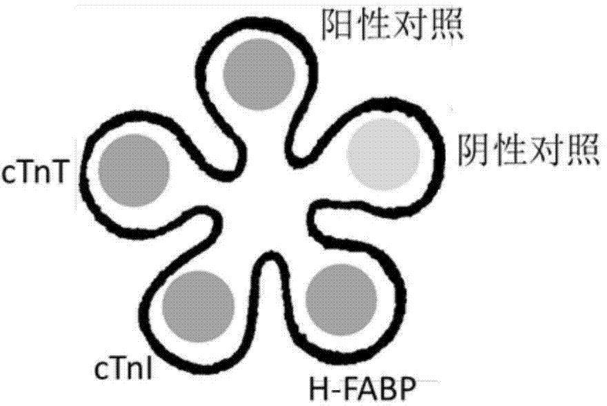
[0047] 1. Making paper-based chips: Using a pair of metal molds that mesh with each other and have sharp edges, the nitrocellulose paper is made into flower-shaped paper-based chips with five petals at room temperature by embossing according to the designed size.

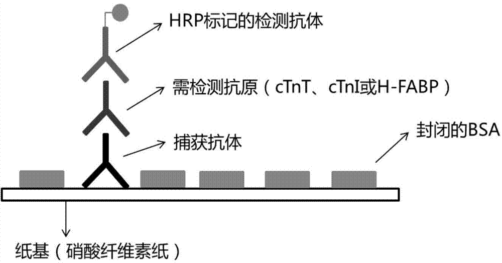
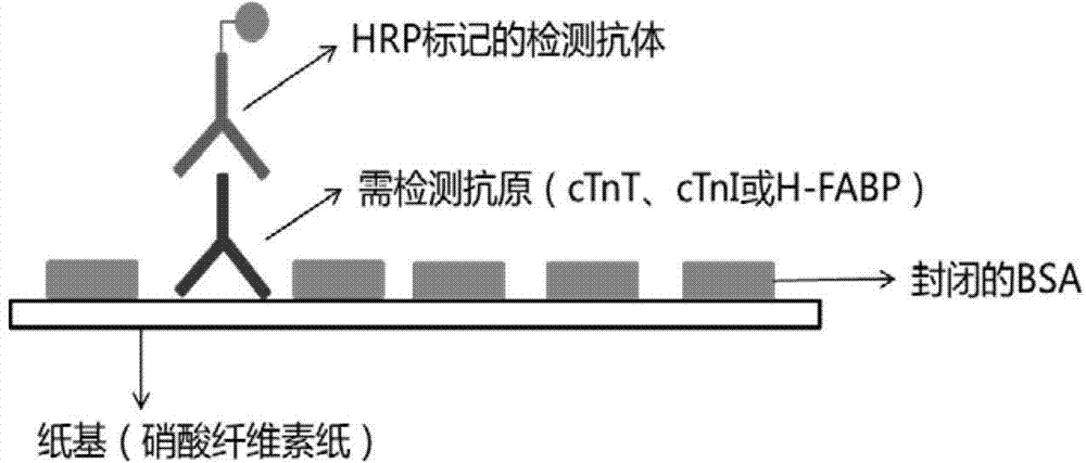
[0048] 2. The distribution of the detection area of ​​the chip is as follows: image 3 shown. Add 1 μL cTnI, cTnT and H-FABP capture antibody dropwise to the cTnI, cTnT and H-FABP detection area, add 1 μL PBS solution dropwise to the negative control detection area, add 1 μL cTnI capture antibody dropwise to the positive control detection area, and dry at room temperature.

[0049] 3. The above dried paper-based chips were sealed with 10% (w / v) BSA solution at room temperature for 30 min....

Embodiment 2

[0051] Embodiment 2: Myocardial infarction rapid detection kit

[0052] The detection kit includes: paper-based chip I prepared in Example 1; cTnI, cTnT and H-FABP detection antibody mixtures labeled with horseradish peroxidase; positive control sample (cTnI standard), sample washing solution 1. Sample washing solution 2 and horseradish peroxidase substrate.

[0053] Sample washing solution 1 is a PBS solution containing 0.05% (v / v) Tween-20 (pH 7.4-7.8); sample washing solution 2 is a PBS solution containing 0.2% (v / v) Tween-20 (pH 7.4-7.8 ).

Embodiment 3

[0054] Example 3: Detection of Paper-based Chip I Clinical Serum Samples

[0055] 1. Source of serum samples: normal and patient serum from the laboratory department of the hospital.

[0056] 2. Sample detection: use the myocardial infarction rapid detection kit in Example 2 for detection. Add 1 μL of serum samples to the cTnI, cTnT, H-FABP and negative control detection areas of the paper-based chip I, and add 1 μL of positive control samples to the positive control detection area. After 2 minutes, put the chip in sample washing solution 1 to wash off excess samples, 3 minutes / 3 times.

[0057] 3. Add 1 μL of the detection antibody mixture dropwise to each detection area and let it stand for 2 minutes to form antibody-antigen-enzyme-labeled antibody complexes. Then put the paper-based chip I into the sample washing solution 2 (PBS solution containing 0.2% v / v Tween-20), 3 min / 3 times.

[0058] 4. Mix the chemiluminescent substrate of horseradish peroxidase (Milipore, Immob...


PUM


Abstract

The invention discloses a rapid detection kit for myocardial infarction. The kit comprises a cardiac troponin I (cTnI) detection reagent, a cardiac troponin T (cTnT) detection reagent and a heart-type fatty acid-binding protein (H-FABP) detection reagent. According to the rapid detection kit disclosed by the invention, three indexes are simultaneously used and are integrated together to prepare the kit, and the kit is convenient to carry and simple in detection process; by simultaneously rapidly detecting cTnI, cTnT and H-FABP, AMI can be diagnosed or eliminated within 1 hour after disease attack, disease attack stages can be determined, the critical illness degree can be evaluated, and the advantages of bedside and on-site rapid applications can be achieved; AMI can be confirmed or eliminated in a very early stage, risk stratification can be achieved, and the purpose of monitoring AMI in the whole course can be achieved; and by virtue of triple detection, a maximum detection time window of AMI can be provided, and high sensitivity and relatively strong specificity (avoiding false positive) can also be achieved.

Description

technical field [0001] The invention relates to the technical field of disease detection preparations, in particular to a rapid detection kit for myocardial infarction. Background technique [0002] Myocardial infarction (MI) is a disease in which coronary artery occlusion and blood flow interruption cause local necrosis of some myocardium due to severe and persistent ischemia. Clinical symptoms include severe and long-lasting retrosternal pain, fever, leukocytosis, accelerated erythrocyte sedimentation rate, increased serum myocardial enzyme activity, and progressive ECG changes. Arrhythmia, shock, or heart failure may occur. Myocardial infarction is a kind of heart disease belonging to coronary atherosclerosis. [0003] Acute myocardial infarction (AMI) refers to the acute necrosis of part of the myocardium caused by persistent and severe myocardial ischemia. At present, the clinical diagnosis of AMI relies on the detection of cardiac markers, electrocardiogram, imaging ...

Claims


Application Information

Patent Timeline
IPC IPC(8): G01N33/68
CPCG01N33/6893G01N2800/324
Inventor 王义明罗国安
Owner 王义明
Why Eureka
Try Eureka