Method for separating and purifying rat dermal microvascular endothelium cells

An endothelial cell, separation and purification technology is applied in the field of in vitro culture of rat dermal microvascular endothelial cells to achieve the effect of improving the recovery rate and high purity

Inactive Publication Date: 2015-09-02
BEIJING UNIV OF AGRI
View PDF2 Cites 5 Cited by
  • Summary
  • Abstract
  • Description
  • Claims
  • Application Information

AI Technical Summary

Problems solved by technology

[0005] The main technical problem of the present invention is to solve the deficiencies of the existing microvascular endothelial cell separation and purification technology, and to provide a method for isolating and cultivating high-purity rat dermal microvascular endothelial cells

Method used


Image

  • Method for separating and purifying rat dermal microvascular endothelium cells
  • Method for separating and purifying rat dermal microvascular endothelium cells
  • Method for separating and purifying rat dermal microvascular endothelium cells

Examples


Embodiment

[0020] 1. Main experimental materials

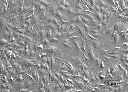
[0021] (1) Experimental animals: 5 3-day-old SD rats.

[0022] (2) Mouse anti-rat CD31 monoclonal antibody: purchased from AbD Serotec Company, product number MCA 1334G, the specification is 200 μL (including 200 μg).

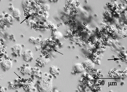
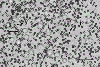
[0023] (3) Mouse IgG magnetic bead kit: including 5 mL immunomagnetic beads coated with anti-mouse IgG, 3 tubes of release buffer component 1 (15000-20000 U freeze-dried DNase per tube ), and 2 mL release buffer component 2.

[0024] Preparation of release buffer: add 0.3mL of release buffer component 2 to each tube of release buffer component 1 to dissolve, aliquot and store in freezer, and add 4 μL per ml of sample when in use.

[0025] (4) 0.01 M PBS: Take 8.0 g of NaCl, Na 2 HPO 4 2.9 g, KH 2 PO 4 0.2 g and 0.2 g KCl, dissolved in 1000 mL ultrapure water, sterilized by 0.22 μm filter, and stored in a refrigerator at 4°C.

[0026] (5) Sample buffer: prepared with 0.01 M PBS, containing 0.1% bovine serum album...


PUM


Abstract

The invention discloses a method for separating and purifying rat dermal microvascular endothelium cells. The method for separating and purifying the rat dermal microvascular endothelium cells relates to the technical field of animal blood vessel endothelium cell culture in vitro, in particular to a method for culturing the rat dermal microvascular endothelium cells in vitro. The method is characterized by the following steps of digesting rat skin through neutral protease II, separating to obtain a dermal layer, digesting through collagenase I and II, obtaining single-cell suspension, and utilizing a CD31 immunomagnetic beads sorting technology to obtain purified target cells. Through the method, the pollution of parenchyma cells in microvascular endothelium cell culture can be simply, conveniently and effectively solved, the rat dermal microvascular endothelium cells with high purity can be obtained through separating, and no obvious morphological feature change occurred after continuous cell culture for 20 times.

Description

technical field [0001] The invention relates to the technical field of mammalian cell culture, in particular to an in vitro culture method of rat dermal microvascular endothelial cells. Background technique [0002] Microvascular endothelial cells occupy key anatomical positions, have a variety of biological functions, and have significant heterogeneity in different tissues or organs. Platelet endothelial cell adhesion molecule 1 (PECAM-1, CD31) is its specificity. surface molecules. The dermis is a tissue located between the skin epidermis and subcutaneous tissue, which is rich in microvessels and is the place for the metabolism and exchange of skin nutrients. The dysfunction of dermal microvascular endothelial cells is the key link in the occurrence of various skin lesions such as skin inflammation, redness, and bleeding. , Rat dermal microvascular endothelial cells cultured in vitro are commonly used models for related research. [0003] The separation of microvessels i...

Claims


Application Information

Patent Timeline
Patent Type & Authority Applications(China)
IPC IPC(8): C12N5/071
Inventor 张涛穆祥董虹胡格杨重锦
Owner BEIJING UNIV OF AGRI
Why Eureka
Try Eureka