A method for separating human amniotic epithelial cells

A technology for amnion epithelial cells and separation methods, which is applied in the direction of embryonic cells, animal cells, vertebrate cells, etc., can solve difficult problems such as amnion epithelial cells, and achieve industrialization, high differentiation potential, and high separation success rate Effect

Active Publication Date: 2018-06-22
CHONGQING CELL BIOENG TECH CO LTD
View PDF4 Cites 2 Cited by
  • Summary
  • Abstract
  • Description
  • Claims
  • Application Information

AI Technical Summary

Problems solved by technology

This makes it difficult to ensure sufficient quantity and purity of amnion epithelial cells for research

Method used


Image

  • A method for separating human amniotic epithelial cells
  • A method for separating human amniotic epithelial cells
  • A method for separating human amniotic epithelial cells

Examples


Embodiment Construction

[0026] The present invention will be further described below in conjunction with the pictures.

[0027] The separation method of described a kind of human amniotic membrane epithelial cell, comprises the following steps:

[0028] The first step is to collect and transport the placenta. First, put the collected fresh placenta into a sterile plastic bag filled with 0.9% mass concentration saline, seal it and put it into a protective box; ~8°C transport box, and transported to the cell separation room within 12 hours; this process ensures the freshness of placental tissue and the activity of amniotic epithelial cells, providing quality assurance for the subsequent isolation and culture.

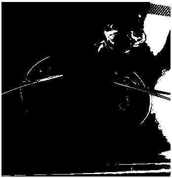
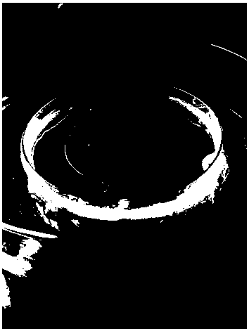
[0029] In the second step, the amniotic membrane is torn out, and the cell separation personnel place the received fresh placenta in the disc, first wash the surface of the fresh placenta with normal saline, and then completely tear off the amniotic membrane from the fresh placenta (see figure ...


PUM


Abstract

The present invention relates to a human amniotic epithelial cell separation method, which comprises: 1, collecting and transporting placenta; 2, tearing and taking the amnion; 3, rinsing the amnion; 4, cutting the amnion; 5, digesting the amnion epithelium surface; 6, terminating the digestion; 7, collecting cells through centrifugation; and 8, resuspending the amniotic epithelial cells, and carrying out inoculating culture. According to the present invention, the high purity amniotic epithelial cells can be separated, the number of the cells is more, the cell activity is high, and the basis is established for the research and the clinical applications of the amniotic epithelial cells.

Description

technical field [0001] The invention relates to a method for separating cells, in particular to a method for separating human amniotic epithelial cells. Background technique [0002] The amniotic membrane is the product of embryonic development. It is a smooth, translucent film without blood vessels and nerves. Its inner side surrounds the embryo, and its outer side is continuous with the chorion. Its thickness is 70-180 um and it has high toughness. As part of the placenta, the amniotic membrane provides the fetus with an adjustable space and suitable environment for local movement. Through filtration and physical buffering, it secretes nutritional factors, maintains the balance of water and electrolytes in the amniotic fluid, and inhibits the mother’s immune response to the embryo. Protect developing embryos. [0003] The amniotic epithelial cells originate from the epiblast on day 8 after fertilization, which is the same as the origin of the final development of the fetu...

Claims


Application Information

Patent Timeline
Patent Type & Authority Patents(China)
IPC IPC(8): C12N5/073
Inventor 潘华锋贾少炼李闯祝文韬王力句凤华徐志国李伟崔涛邓人伟杨娅毛敏吴金丹旷亚丽甘兵郭雪芹李廷玉符州
Owner CHONGQING CELL BIOENG TECH CO LTD
Why Eureka
Try Eureka