Preparation method of transmission electron microscope sample of paraffin-embedded section tissue

A transmission electron microscope sample and paraffin-embedded technology, which is applied to the sample preparation of paraffin-embedded sliced ​​tissue and the field of transmission electron microscope sample preparation of paraffin-embedded sliced ​​tissue, can solve the problem that the embedded block cannot be completely peeled off, environmental and health effects, Problems such as too large embedding surface can reduce the impact on the environment and health, save the amount of use, and reduce degradation and deformation.

Active Publication Date: 2015-10-14
GUANGXI MEDICAL UNIVERSITY
View PDF6 Cites 19 Cited by
  • Summary
  • Abstract
  • Description
  • Claims
  • Application Information

AI Technical Summary

Problems solved by technology

The dewaxing, fixation, dehydration, soaking, and embedding operations of paraffin-embedded tissue TEM sample preparation involve a variety of toxic and volatile liquids such as xylene, glutaraldehyde, osmic acid, acetone, etc. The open operation on the glass slide will not only change the concentration due to the volatilization of the liquid, which will affect the effect of dewaxing, fixing, dehydrati

Method used


Image

  • Preparation method of transmission electron microscope sample of paraffin-embedded section tissue
  • Preparation method of transmission electron microscope sample of paraffin-embedded section tissue
  • Preparation method of transmission electron microscope sample of paraffin-embedded section tissue

Examples


Embodiment 1

[0048] Preparation of the EP tube isolation ring: cut off the EP tube nozzle and a complete circle of tube below it as the isolation ring for later use.

[0049] The specific operation is as follows: cut off the mouth of the 0.5mL conical EP tube and a complete circle of tube with a length of 1 mm below it to obtain the isolation ring of the EP tube.

Embodiment 2

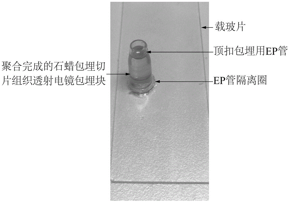
[0051] Preparation of EP tube for top buckle embedding: continue to cut off a complete circle of tube wall from the EP tube prepared after the EP tube isolation ring, and cut off the bottom tip of the EP tube, and the remaining tube is used as EP for top buckle embedding. The tube is ready for use, and the bottom tip of the cut EP tube is used for the EP tube for final sealing and embedding.

[0052] The specific operation is as follows: After cutting off the mouth of the 0.5mL conical EP tube and a complete circle of tube with a length of 1mm below it, continue to cut off the complete circle of tube wall until the remaining tube can just fit into the 0.5mL conical EP tube. Cut off the tip of the bottom of the EP tube in the EP tube isolation circle prepared by the shaped EP tube, and finally obtain the EP tube for top buckle embedding, and cut off the bottom tip of the EP tube for final sealing of the EP tube for top buckle embedding.

Embodiment 3

[0054] A transmission electron microscope sample preparation method for paraffin-embedded section tissue, consisting of the following steps in sequence,

[0055] 1. Adjust the laboratory temperature to 26°C and humidity to 45%;

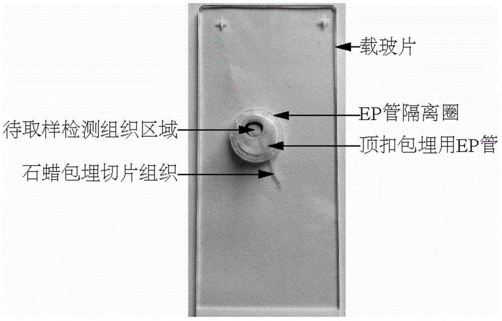
[0056] 2. Provide a glass slide with renal biopsy paraffin-embedded section tissue. The front side of the glass slide has the renal biopsy paraffin-embedded section tissue of the area to be sampled. The area to be sampled contains the renal biopsy tissue to be tested, and the sample is to be sampled using an inverted microscope. Preliminary positioning of the detected renal biopsy tissue;

[0057] 3. Preparation of various pure embedding agents (chemical reagents used are all analytical pure):

[0058] 1) Preparation of 618 embedding agent: Add 12 parts of 618, 8 parts of DDSA, 1 part of DBP, and 0.5 parts of DMP-30 into the measuring cup one by one according to the parts by weight, stir thoroughly for 60 minutes, and place in a vacuum drying oven S...


PUM

PropertyMeasurementUnit
Lengthaaaaaaaaaa

Abstract

The invention discloses a preparation method of a transmission electron microscope sample of a paraffin-embedded section tissue. The preparation method comprises the steps of (1) regulating the temperature of a laboratory to be 22-30 DEG C, and regulating the humidity of the laboratory to be smaller than or equal to 45%; (2) providing a glass slide with the paraffin-embedded section tissue; (3) preparing various pure embedding mediums; (4) preparing isolating rings of EP pipes; (5) preparing EP pipes for top buckling and embedding or prefabricated embedding blocks; (6) dewaxing and hydrating; (7) arranging the isolating rings of the EP pipes; (8) fixing and rinsing; (9) dehydrating and soaking; (10) carrying out top buckling and embedding; (11) polymerizing; (12) stripping; and (13) repairing, slicing, dyeing as well as observing and collecting images by an electron microscope. The preparation method has the beneficial effects that the amount of reagents can be saved, the errors caused by the volatilization of the reagents can be reduced, and meanwhile, the obtained sample can be completely stripped from the glass slide, so that the ultra microstructure of the paraffin-embedded section tissue can be further observed and researched conveniently.

Description

technical field [0001] The invention belongs to the technical field of experimental sample processing, and relates to a sample preparation method for paraffin-embedded section tissue, in particular to a transmission electron microscope sample preparation method for paraffin-embedded section tissue. Background technique [0002] Paraffin-embedded section technology is an important method in the field of biomedical research, especially in clinical pathological diagnosis. By staining the sections of paraffin-embedded tissues, the morphological changes in the tissues can be observed, thereby assisting in the diagnosis of diseases and clarifying the etiology. [0003] Generally, the thickness of paraffin-embedded tissue slices is mostly 2-5 μm. After the slices are sliced, they are spread on glass slides for further HE staining or immunohistochemical staining. Different structures are used to present different colors to facilitate observation and judgment. Organizational structu...

Claims


Application Information

Patent Timeline
IPC IPC(8): G01N1/28
Inventor 黄远洁莫肖敏孟春梅成晓静李卫东郑华
Owner GUANGXI MEDICAL UNIVERSITY
Why Eureka
Try Eureka