Immunomagnetic bead for sorting human cells and preparation and cutting-off method of immunomagnetic bead

A technology of magnetic beads and cells, which is applied in the field of medical biology experiments, can solve the problems of cell waste, unfavorable culture, and long time consumption, and achieve the effect of high excision efficiency

Active Publication Date: 2015-11-25
SHANGHAI BAIZE MEDICAL APP & INSTR CO LTD +2
View PDF3 Cites 29 Cited by
  • Summary
  • Abstract
  • Description
  • Claims
  • Application Information

AI Technical Summary

Problems solved by technology

However, because this method takes a long time, it is not conducive to subsequent cell culture, and the purity of th

Method used


Image

  • Immunomagnetic bead for sorting human cells and preparation and cutting-off method of immunomagnetic bead
  • Immunomagnetic bead for sorting human cells and preparation and cutting-off method of immunomagnetic bead
  • Immunomagnetic bead for sorting human cells and preparation and cutting-off method of immunomagnetic bead

Examples


Example Embodiment

[0114] Example 1: Preparation of magnetic bead-nucleic acid-antibody complex

[0115] [1] Take the magnetic beads (0.5 mg) with amino groups on the surface into a 1.5 ml EP tube, magnetically separate, and discard the supernatant.

[0116] [2] Resuspend the magnetic bead pellets in 1 ml of PBS buffer, then magnetically separate, and discard the supernatant.

[0117] [3] Repeat step 2 two or three times.

[0118] [4] Add 600-800ul of coupling agent glutaraldehyde, place it in a mixing rotator and incubate at room temperature for 1.5h.

[0119] [5] Take the nucleic acid fragment (SEQ ID NO: 1) (magnetic beads: 1 mg: 50 ug of nucleic acid) into a 1.5 ml centrifuge tube, add 600-800 ul of glutaraldehyde, and place it on a mixing rotator and incubate at room temperature for 1.5 hours.

[0120] [6] Remove the incubated magnetic beads from the rotator, magnetically separate, and remove the supernatant.

[0121] [7] Add the nucleic acid fragments after the incubation in step 4, resuspend the magn...

Example Embodiment

[0129] Example 2: Magnetic bead-nucleic acid-antibody complex cell sorting method

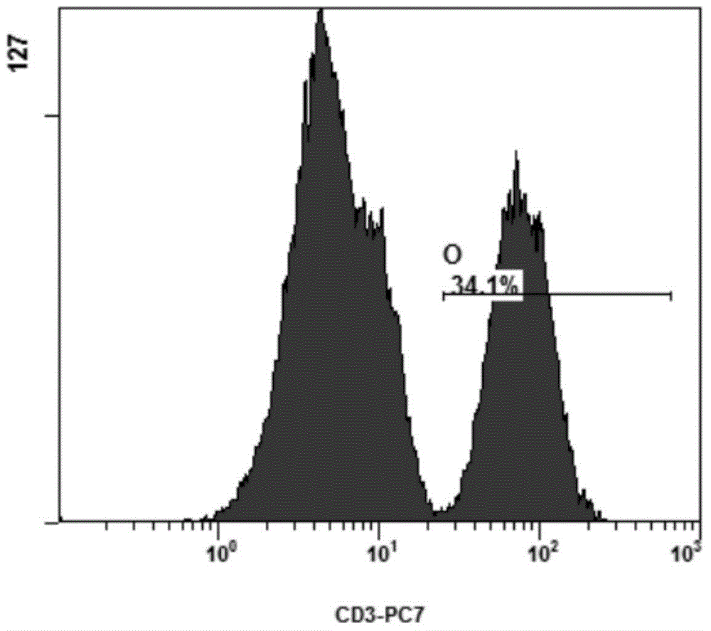
[0130] [1] Take 1ml of human peripheral blood mononuclear cells (PBMC) and centrifuge at 1500rpm for 5min. figure 1 Flow cytometry showing the proportion of CD3-PECy7 positive for peripheral blood mononuclear cells (initial cell number 1×10 8 , The proportion of CD3 positive cells is 34.1%, that is, containing 3.41×10 7 CD3 positive cells).

[0131] [2] Discard the supernatant, add PBS to resuspend the cells, and centrifuge at 1500 rpm for 5 min.

[0132] [3] Repeat step 2 twice.

[0133] [4] Resuspend the cells with a small amount of PBS, add the magnetic beads prepared in Example 1, mix them evenly, and place them in a mixing rotator to incubate at room temperature for 1 hour.

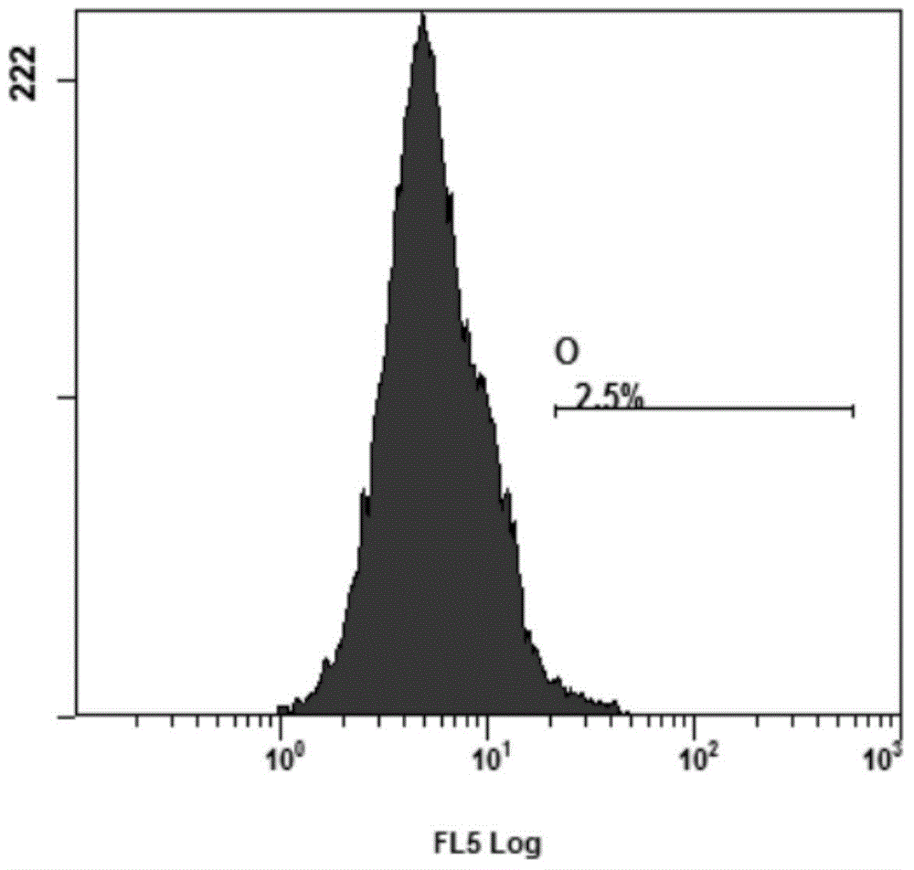
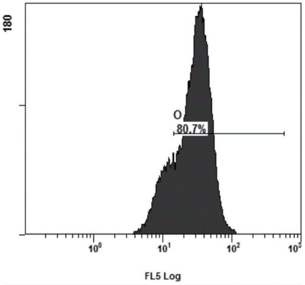
[0134] [5] After the incubation, the cells are magnetically separated, and the supernatant is aspirated to be negative cells. figure 2 After the sorting is completed, the proportion of CD3-PECy7 positive cells detected by flow...

Example Embodiment

[0138] Embodiment 3: Magnetic bead removal method

[0139] [1] The positive cells collected in Example 2 were centrifuged at low speed.

[0140] [2] Discard the supernatant, add 90ul double enzyme digestion buffer to resuspend the cells.

[0141] [3] Add enzyme and bath in 37℃ water for 10 minutes.

[0142] [4] Magnetic separation, aspirate the supernatant to a new centrifuge tube.

[0143] [5] Centrifuge, discard the supernatant, and resuspend in a small amount of PBS to form the positive cells without magnetic beads after sorting.

[0144] Table 1 below shows the magnetic bead excision experiments using different enzyme digestion methods, and the measured excision time, excision efficiency and cell activity.

[0145] Table 1

[0146]

[0147] 1: The time it takes to completely remove the magnetic beads, this experiment is to recover 3.1×10 7 Each cell shall prevail.

[0148] 2: Live and dead cells are counted by staining with trypan blue and then counting under a microscope.

[0149] 3: Ce...


PUM


Abstract

The invention provides an immunomagnetic bead for sorting human cells and a preparation and cutting-off method of the immunomagnetic bead and particularly relates to a magnetic bead-nucleic acid-antibody compound or a kit containing the magnetic bead-nucleic acid-antibody compound. The compound comprises a magnetic bead, nucleic acid covalently coupled to the magnetic bead and an antibody covalently coupled with the nucleic acid, wherein the nucleic acid comprises at least one enzyme digestion site, and the antibody can specifically bind with antigen on the surfaces of to-be-screened cells. The invention further comprises the purpose and preparation method of the magnetic bead-nucleic acid-antibody compound and a method for using the magnetic bead-nucleic acid-antibody compound to perform cell sorting.

Description

Technical field [0001] The invention belongs to the technical field of medical biology experiments, and relates to an immunomagnetic bead for sorting human cells, and a preparation and excision method thereof. Background technique [0002] Tumors are one of the diseases that seriously endanger human health. Traditional treatment methods such as surgery, radiotherapy, and chemotherapy have been difficult to kill the remaining tumor cells in the body. In the 21st century, with the technological advancement of tumor treatment, it has developed into the technical system of biological treatment. Among them, biological therapy is currently the most mature and widely used tumor biological therapy method. Biological therapy is to collect mononuclear cells (PBMC) from the patient's peripheral blood, and then send them to the GMP laboratory for culture, expansion, induction, and tumor antigen stimulation, so as to obtain DC cells that can recognize cancer cells and have high tumor killing...

Claims


Application Information

Patent Timeline
IPC IPC(8): C12Q1/68
CPCC12Q1/6804C12Q2521/301
Inventor 钱其军陆元修杨华宋启平李林芳师传胤
Owner SHANGHAI BAIZE MEDICAL APP & INSTR CO LTD
Why Eureka
Try Eureka