A transuterine fallopian tube ultrasound detection method, diagnostic instrument and transducer

A technology of ultrasonic transducer and ultrasonic diagnostic instrument, which is applied in the direction of catheter, surgery, etc., can solve the problems of low imaging resolution, small imaging field of view, detection, etc., achieve high imaging resolution, strong penetrating ability, and improve the clarity of pictures degree of effect

Active Publication Date: 2018-03-13
上海爱声生物医疗科技有限公司
View PDF4 Cites 0 Cited by
  • Summary
  • Abstract
  • Description
  • Claims
  • Application Information

AI Technical Summary

Problems solved by technology

It consists of a flexible imaging optical fiber, which can be inserted into the fallopian tube to perform optical imaging on the internal structure and obstruction of the entire fallopian tube, but the imaging field of view is small, the imaging resolution is low (millimeter level), and it can only image the surface of the tissue. Inability to observe deep tissue lesions
[0007] There are certain shortcomings in the three existing technologies. Laparoscopy and hysteroscopy can only observe from the outside of the fallopian tube. Although the fallopian tube can observe the inside of the fallopian tube, it can only image the tissue surface and cannot observe deep tissue lesions
Therefore, the current fallopian tube diagnostic technology cannot comprehensively and effectively detect the fallopian tube

Method used


Image

  • A transuterine fallopian tube ultrasound detection method, diagnostic instrument and transducer
  • A transuterine fallopian tube ultrasound detection method, diagnostic instrument and transducer
  • A transuterine fallopian tube ultrasound detection method, diagnostic instrument and transducer

Examples


Embodiment 1

[0075] Example 1: Intrafallopian Tube Ultrasound Transducer Using Single Beam Technology

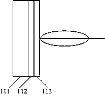
[0076] combine figure 1 , This embodiment describes in detail the intrafallopian ultrasound transducer using single beam technology, which includes an ultrasound transducer unit composed of a backing layer 111 , a piezoelectric layer 112 and an acoustic matching layer 113 that are closely connected in sequence. Correspondingly, the ultrasonic catheter driving its movement drives its 360-degree rotation.

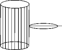
[0077] In different embodiments, the ultrasonic transducer in the fallopian tube can also be a cylindrical array ultrasonic transducer, which includes a plurality of ultrasonic transducer units distributed 360 degrees along the cylindrical surface, such as figure 2 shown. Correspondingly, the moving ultrasonic catheter only needs to be driven to move back and forth, and does not need to be rotated.

[0078] The ultrasonic transducer in the fallopian tube of this embodiment can enter...

Embodiment 2

[0080] Example 2: Ultrasound Focusing Transducer in the Fallopian Tube Using Whole Acoustic Structure Focusing Technology

[0081] like Figure 4 Shown is a schematic diagram of the intrafallopian tube ultrasonic focusing transducer of this embodiment, which includes a backing layer 111, a piezoelectric layer 112 and an acoustic matching layer 113 that are closely connected in sequence, wherein: the backing layer 111, the piezoelectric layer 112 and The acoustic matching layers 113 all have mechanical curved surfaces, and the radii of curvature of the three can be calculated and set according to the requirements of the focused sound field. The focus factor K is defined as the ratio of the focal length f to the transducer aperture d, ie: K=f / d. Given the focus factor K and the focal length f, the size of the aperture d can be calculated.

Embodiment 3

[0082] Embodiment 3: Intrafallopian tube ultrasonic focusing transducer using acoustic lens focusing technology

[0083] like Figure 5 Shown is a schematic diagram of the intrafallopian tube ultrasonic focusing transducer of this embodiment, which includes a backing layer 111, a piezoelectric layer 112, an acoustic matching layer 113, and an acoustic lens 114 that are tightly connected in sequence, wherein the acoustic lens 4 has a mechanically curved surface , its radius of curvature can be calculated and set according to the requirements of the focused sound field.

[0084] The acoustic lens 114 can be a plano-convex lens or a plano-concave lens, which is determined according to the sound velocity of the lens material. For lens materials whose sound velocity is lower than that of the medium, it is a plano-convex lens, such as Figure 6 Shown by the dotted line in the middle; for the lens material whose sound velocity is higher than the sound velocity of the medium, it is ...


PUM


Abstract

The invention discloses a transuterine fallopian tube ultrasound detection method, a diagnostic instrument and a transducer. The method comprises: passing an ultrasound transducer in the fallopian tube with a center frequency of 10 MHz to 100 MHz through the uterus through an ultrasonic catheter with a diameter of 0.5 mm to 5 mm. Send it into the fallopian tube to be tested; transmit and receive ultrasonic signals 360 degrees to the fallopian tube to be tested; and withdraw the ultrasound transducer in the fallopian tube at the same time. The diagnostic instrument includes: an ultrasound catheter with a diameter of 0.5mm~5mm, an intra-fallopian ultrasound transducer with a center frequency of 10MHz~100MHz is installed at the front end, and the back end is connected with a retraction / driving device; the retraction / driving device is connected with an electronic imaging system . The transducer includes: an ultrasonic transducer unit composed of a backing layer, a piezoelectric layer and an acoustic matching layer closely connected in sequence. The invention sends the ultrasonic transducer into the fallopian tube through the uterus, improves the imaging resolution, has strong penetration rate, and has no blind angle of vision.

Description

technical field [0001] The invention relates to a fallopian tube diagnostic instrument, in particular to a transuterine fallopian tube ultrasonic detection method, a diagnostic instrument and a transducer. Background technique [0002] Blocked fallopian tubes are the leading cause of female infertility, accounting for about 25% to 35%. The fallopian tube is the channel connecting the ovary and the uterus. It is responsible for the function of fallopian, egg storage, insemination, providing a place for the combination of sperm and egg, and transporting the fertilized egg to the endometrium. If the fallopian tubes are blocked, a series of conception processes such as the combination of sperm and eggs will be hindered, resulting in female infertility. The fallopian tubes are located on both sides of the uterus. There are two openings on each side of the fallopian tubes. The inner end opens in the uterine cavity and connects with the outer corner of the uterine fundus, and the ...

Claims


Application Information

Patent Timeline
Patent Type & Authority Patents(China)
IPC IPC(8): A61B8/12
Inventor 袁建人李翔陈友伟
Owner 上海爱声生物医疗科技有限公司
Why Eureka
Try Eureka