Growth matrices for stem cell propagation in vitro and in tissue regeneration

A stem cell and tissue technology, applied in the field of culturing stem cells

Inactive Publication Date: 2015-12-09
RUTGERS THE STATE UNIV +1
View PDF4 Cites 2 Cited by
  • Summary
  • Abstract
  • Description
  • Claims
  • Application Information

AI Technical Summary

Problems solved by technology

[0006] Stem cell therapy is a promising field for tissue engineering and regeneration, but it has shown on

Method used


Image

  • Growth matrices for stem cell propagation in vitro and in tissue regeneration
  • Growth matrices for stem cell propagation in vitro and in tissue regeneration
  • Growth matrices for stem cell propagation in vitro and in tissue regeneration

Examples


Embodiment 1

[0048] Embodiment 1 (stem cell morphology, proliferation and differentiation)

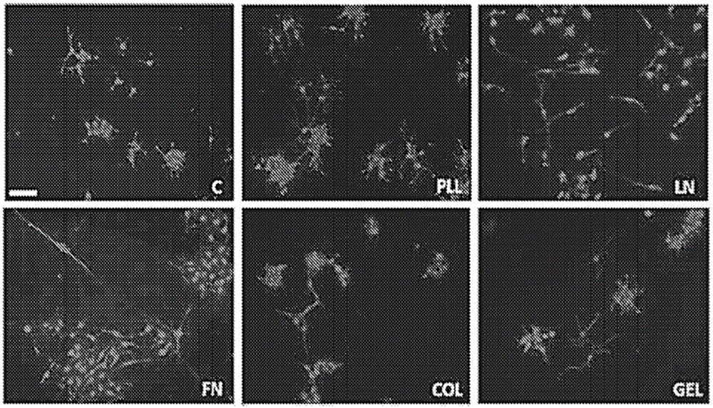
[0049] Neural stem cells: Two cell types were used to assess the functionality of modified chitosan membranes and microspheres: the immortalized cell line RG3.6, which is positive for green fluorescent protein (GFP) by day 13.5 embryos + ) from rat neocortical cells; and primary NSCs harvested from day 13.5 embryonic EGFP(SD-Tg(GFP)Bal / 2Rrrc(RRRC:0065)) rat neocortex. For specific experiments, the RG3.6 cell line was used instead of primary cells to eliminate multiple variables seen with heterogeneous primary cell cultures and to achieve greater consistency in matrix optimization. RG3.6 and primary NSCs were maintained supplemented with B27, gentamicin (50 μg / mL), apotransferrin (50 μg / mL), and rhFGF-2 (10 ng / mL + 1 ng / mL heparan sulfate ) in DMEM / F12 medium. They were grown as neurospheres or as attached monolayers on polyornithine / fibronectin-coated petri dishes.

[0050] Chitosan film: A 3% w...

Embodiment 2

[0055] Embodiment 2 (stent efficiency)

[0056] The biological activity of human fibroblast growth factor-2 (hFGF-2) bound to scaffolds was verified by measuring cell growth using MTT assay and analyzing the morphology of NSCs. Heparin sodium salt from bovine intestinal mucosa was purchased from Sigma (St Louis, MO). Recombinant human fibroblast growth factor-2 (rhFGF-2) was purchased from Peprotech (Rocky Hill, NJ). Genipin was purchased from Wako Pure Chemical Industries, Ltd. (Osaka, Japan).

[0057] Cell Growth: Coat 96-well plates with 50 μL of 3% chitosan and air dry overnight. Selection coated wells were incubated with heparin (0.5 mg / mL) and genipin (0.45 mM) overnight at room temperature. Heparin-genipin solutions were prepared in 50 mM HEPES + 0.9% NaCl solution (HBS) (VWR; West Chester, PA). Other wells were incubated overnight in HBS only. The following day, fibronectin (10 ug / mL) was added to each well for 4 hours at 37°C to enhance cell attachment to the chi...

Embodiment 3

[0063] Embodiment 3 (characterization of 3-dimensional support)

[0064] Preparation of chitosan microspheres: Chitosan powder (1.5 g) was dispersed in 50 mL of water containing 2.0% v / v acetic acid to create a 3% chitosan solution. The chitosan solution was stirred mechanically at 700 rpm until completely dissolved. The resulting solution was collected and centrifuged at 2,000 rpm for 10 minutes. Subsequently, the supernatant was collected and the remaining impurities precipitated were discarded. Squeeze the acidic chitosan solution into an alkaline coagulation bath (composed of 2.5 M NaOH:methanol:water (20:30:50 v / v)) using a syringe at a flow rate of 5 mL / hr to form chitosan Microspheres. In order to reduce the surface tension on the needle tip, thereby reducing the size of the microspheres to the desired range, a current of 25 kV was applied. Next, filter the spheres using a 100 μm filter to remove any oversized spheres. Remove it from the ionic solution and rinse 4 ...


PUM


Abstract

The present invention provides a multifunctional 2-D and 3-D matrix for propagation of stein cells, in particular, a cliitosatv-based biomaieria! scaffold is engineered to promote CNS regeneration from primitive neural precursors by stabilizing a recombinant protein, fibroblast growth factor to preserve the cardinal properties of stem cells. The matrix, is further modified by the addition of either the extracellular matrix protein fibronectiii or the small peptide RGD or IK.VAV. A method to manu&eture an injectable multifunctional microsphere scaffold is also disclosed that is suitable as a vehicle for cell transplantation to repair traumatic brain injuries.

Description

[0001] Cross References to Related Applications [0002] This application claims the benefit of U.S. Provisional Application No. 61 / 757,378, filed January 28, 2013, under 35 U.S.C. 119(e), the contents of which are incorporated herein in their entirety. [0003] Statement of Government Support [0004] This invention was made with government support under contracts 09-3207-BIR-E-2 and CBIR12FEL025 awarded by the Brain Injury Research Council of New Jersey. technical field [0005] Generally, the present invention relates to the field of tissue engineering. In particular, the present invention relates to the stabilization of a recombinant protein, fibroblast growth factor-2 (FGF-2), to promote its biological activity in stem cells, and methods for culturing stem cells. Background technique [0006] Stem cell therapy is a promising field of tissue engineering and regeneration, but it has shown only limited success in repairing the central nervous system (CNS), especially the...

Claims


Application Information

Patent Timeline
IPC IPC(8): C12N5/00
CPCA61K35/28A61K35/51A61K35/545C12N5/0068C12N5/0623C12N2501/115C12N2533/52C12N2533/72A61K9/0085A61K9/5078A61K35/30
Inventor S·利维森N·什科普F·卡尔德隆C·赵C·甘地
Owner RUTGERS THE STATE UNIV
Why Eureka
Try Eureka