Application of AcSDKP in limbal stem cell separation culture and method for culturing limbal stem cell

A corneal limbal stem cell, separation and culture technology, applied in the field of corneal limbal stem cell culture, can solve the problems of reduced corneal limbal stem cell purity, affecting the corneal limbal stem cell proliferation, etc., and achieves a good effect of maintaining stemness

Inactive Publication Date: 2016-01-13
GUANGZHOU SALIAI STEMCELL SCI & TECH CO LTD
View PDF0 Cites 4 Cited by
  • Summary
  • Abstract
  • Description
  • Claims
  • Application Information

AI Technical Summary

Problems solved by technology

Fibroblasts will affect the proliferation of limbal stem cells during the later culture process, and the products secreted by them will promote the proliferation a...

Method used


Image

  • Application of AcSDKP in limbal stem cell separation culture and method for culturing limbal stem cell
  • Application of AcSDKP in limbal stem cell separation culture and method for culturing limbal stem cell

Examples


Embodiment 1

[0043] Medium: 90% DMEM medium + 10% FBS, wherein the mass fraction of AcSDKP is 0.5%.

[0044] The eyeballs of dead fetuses with a gestational age of 3-7 months were soaked in PBS containing double-antibody (penicillin-streptomycin, 1×) for 3 minutes in a sterile environment, and then rinsed with PBS without double-antibody.

[0045] Under the operating microscope, use tissue forceps and corneal scissors to cut the human corneal limbal tissue, and then use scissors to cut the tissue into about 1mm 3 blocky in size.

[0046] According to the volume of the tissue block, every 1cm 2 Add 5ml of trypsin with a mass fraction of 0.25% (prepared in PBS solution, pH 7.2-7.4) to the tissue block, digest at 37°C for 15 minutes, add PBS containing 10% FBS to each 1ml of digestion solution to stop digestion, and filter through a 100um mesh sieve , centrifuged at 1000rpm for 5min, discarded the supernatant, added the culture medium to 1×10 5 The density of cell / ml was inoculated into a ...

Embodiment 2

[0049] Medium: 90% DMEM medium + 10% FBS, wherein the mass fraction of AcSDKP is 1%.

[0050] The eyeballs of dead fetuses with a gestational age of 3-7 months were soaked in PBS containing double-antibody (penicillin-streptomycin, 1×) for 3 minutes in a sterile environment, and then rinsed with PBS without double-antibody.

[0051] Under the operating microscope, use tissue forceps and corneal scissors to cut the human corneal limbal tissue, and then use scissors to cut the tissue into about 1mm 3 blocky in size.

[0052] According to the volume of the tissue block, every 1cm 2 Add 5ml of trypsin with a mass fraction of 0.25% (prepared in PBS solution, pH 7.2-7.4) to the tissue block, digest at 37°C for 15 minutes, add PBS containing 10% FBS to each 1ml of digestion solution to stop digestion, and filter through a 100um mesh sieve , centrifuged at 1000rpm for 5min, discarded the supernatant, added the culture medium to 1×10 5 The density of cell / ml was inoculated into a pe...

Embodiment 3

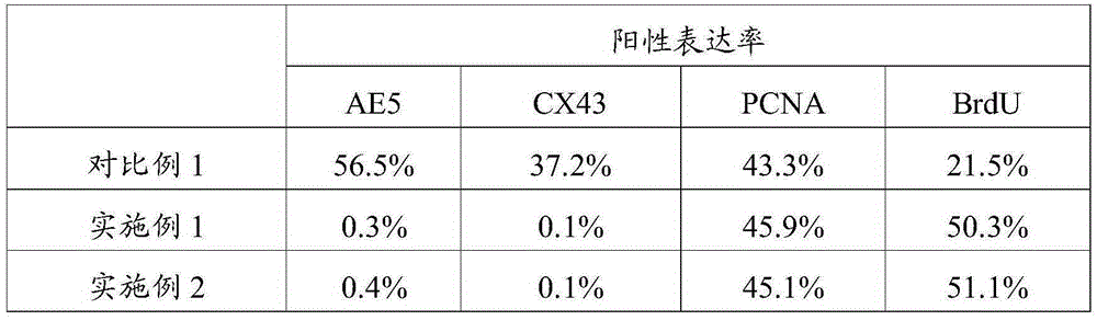
[0061] Flow cytometric detection was performed on the P1 generation cells cultured in Examples 1-2 and Comparative Example 1. The method is:

[0062] 1. Three parallel tests were carried out for each sample, and 1×10 6 The cells were evenly divided into 2 EP tubes, one tube was used as a control, centrifuged at 1500rpm / min for 5min, and the supernatant was discarded.

[0063] 2. Add 1 mL of staining buffer (PBS containing 10% FBS) to resuspend the cells, centrifuge at 1500 rpm / min for 5 min, and discard the supernatant. Repeat the above steps 1 time.

[0064] 3. After discarding the supernatant, add 200 μL of staining buffer to each tube to resuspend, and add 2 μL of specific recognition basic keratin antibody (AE5), connexin antibody (CX43 antibody), and DNA polymerase δ in the sample group. Protein antibody (PCNA) and BrdU antibody (kits used in the experiment, respectively Monoclonal Anti-Connexin43 (Cx43) kit of AlphaDiagnoesticInternational; PEanti-humanPCNA kit of Bio...


PUM


Abstract

The invention relates to the field of stem cell culture, and in particular relates to application of AcSDKP in limbal stem cell separation culture and a method for culturing limbal stem cells. According to the culture medium provided by the invention, fetal calf serum is adopted to promote growth of limbal stem cells, and AcSDKP is adopted to inhibit growth of fibroblast. Therefore, by adopting the culture medium provided by the invention, limbal stem cells can be selectively separated, the purity of the limbal stem cells obtained through separation culture is improved, the number of the limbal stem cells obtained through separation culture is increased, and as growth of fibroblast is inhibited, the limbal stem cells are slightly affected by fibroblast secreta, and are in good dryness state. The experiment shows that when limbal stem cells are separated by using the culture medium provided by the invention, the positive rate of an antibody CX43 in cells is 0.1%, the positive rate of AE5 is 0.3%, the positive rate of BrdU is 50.3%, and the positive rate of PCNA is 45.9%. The effect is remarkably superior to that of the prior art.

Description

technical field [0001] The invention relates to the field of stem cell culture, in particular to the application of AcSDKP in the isolation and culture of limbal stem cells and the culture method of limbal stem cells. Background technique [0002] The corneal limbus is the junction of the cornea, conjunctiva and sclera. The distinguishing mark from the cornea is the termination of Bowman's membrane; the distinguishing mark from the conjunctiva is that it does not contain goblet cells and is about 1 mm wide. There are only epithelial and stroma layers here. The epithelial cell layer is more than 10 layers, arranged irregularly, the cells are small cylindrical, and the nucleus is deeply stained. The deep basal cells are a layer of small cylindrical or cuboid cells, and the nuclei are oval and parallel to the surface. In the basal The papillae are formed in the papilla, forming a special "palisade"-like epithelial structure, which contains pigment and rich vascular network, and...

Claims


Application Information

Patent Timeline
IPC IPC(8): C12N5/074
Inventor 葛啸虎陈海佳王一飞吴子杰张维敏
Owner GUANGZHOU SALIAI STEMCELL SCI & TECH CO LTD
Why Eureka
Try Eureka