Method for establishing hepatocellular carcinoma cis-platinum drug-resisting cell strain
A technology for drug-resistant cells and hepatocellular carcinoma, applied in the field of biomedicine, can solve the problems of poor prognosis of liver cancer patients, and achieve the effect of simple and easy method, high scientific research and production application value
- Summary
- Abstract
- Description
- Claims
- Application Information
AI Technical Summary
Problems solved by technology
Method used
Image
Examples
Embodiment 1
[0034] Method for Inducing and Establishing Liver Cancer Drug-resistant Strain LM3 / CDDP Cells
[0035] (1) Take LM3 cells and use DMEM culture medium after recovery, at 37°C, 5% CO 2 Cultivated in an incubator;
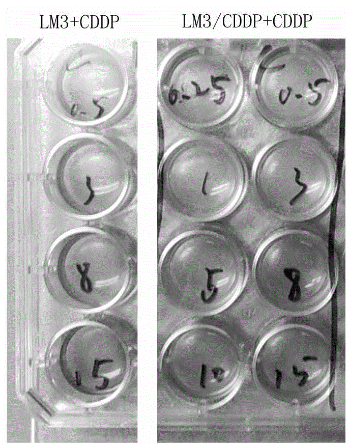
[0036] (2) In order to find the most suitable stimulating concentration of cisplatin, LM3 cells in the logarithmic growth phase were planted in 24-well plates, and 0.1 μg / L, 0.25 μg / L, 0.5 μg / L, 1 μg / L, 2 μg / L , 5μg / L, 10μg / L cisplatin, timely subculture or change medium. Each concentration of chemotherapeutic drugs was added correspondingly at each subculture or medium change. The optimal concentration is the maximum concentration at which the cells can survive and grow slowly after the chemotherapeutic drug is added. The long-term growth referred to in this example refers to the growth time of more than 3 months.
[0037] The experimental results show that: when the concentration of cisplatin is 0.5 μg / mL, LM3 cells can survive but grow very slowly; when the con...
Embodiment 2
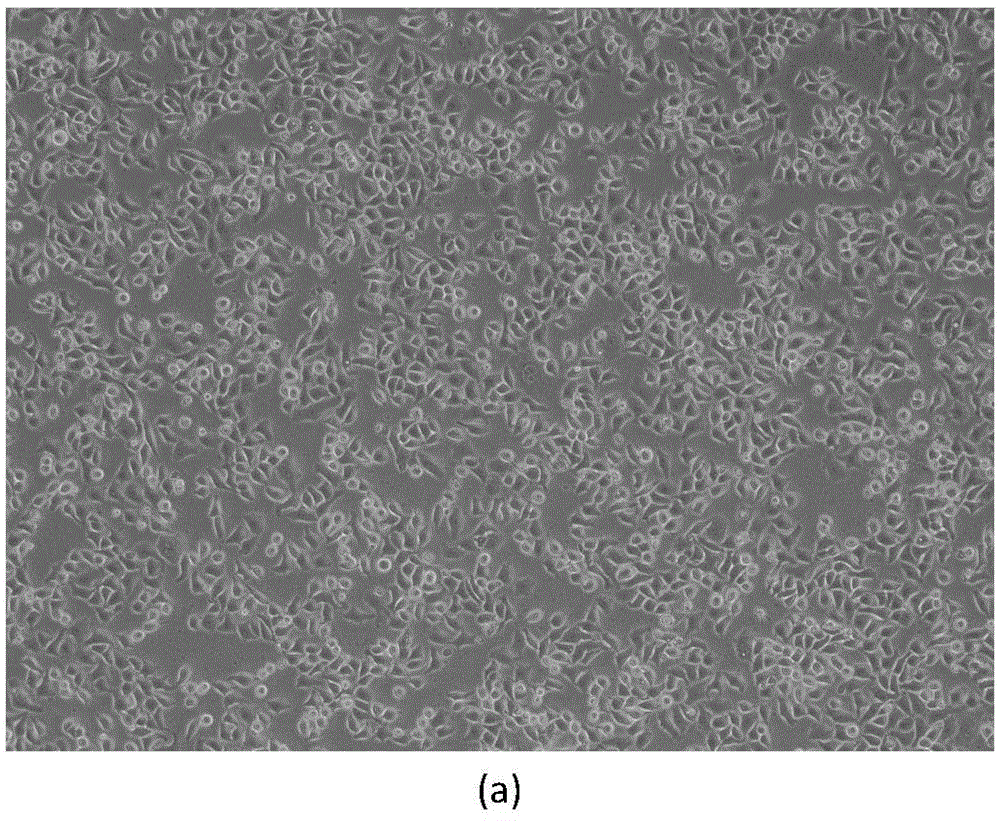
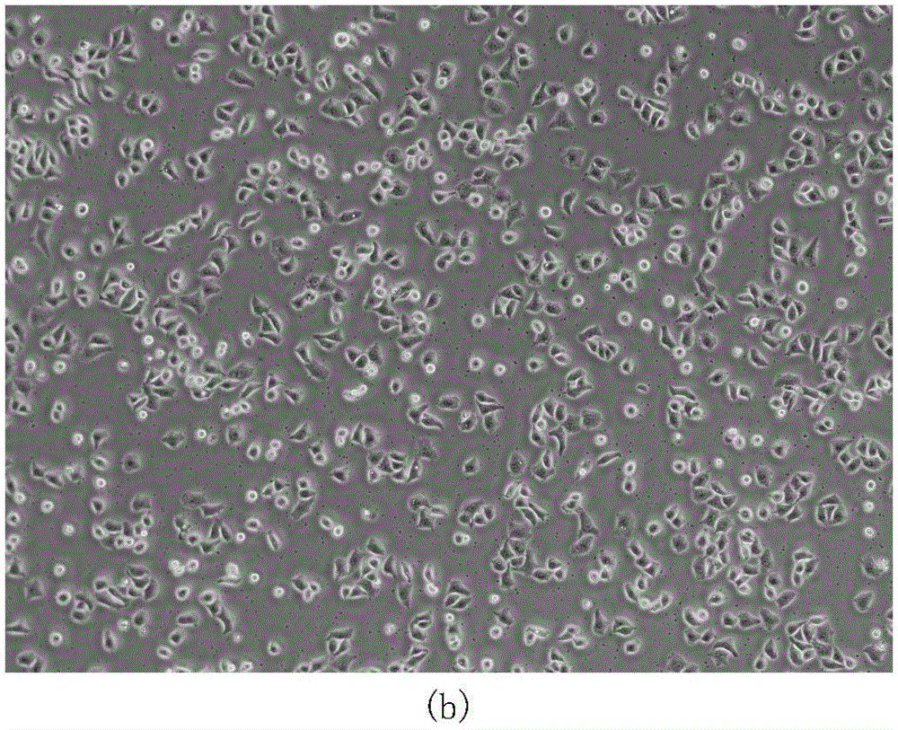
[0040] Morphological Observation of Drug-resistant Liver Cancer LM3 / CDDP Cells
[0041] Use an inverted phase-contrast microscope to observe the morphology of living cells: take LM3 cells and LM3 / CDDP cells in the logarithmic growth phase, wash and replace with normal saline, and observe the morphology of living cells under an inverted microscope with a magnification of 200 times. Such as figure 2 As shown in (a), it can be observed by light microscopy that the LM3 cells are basically the same size, polypodia-like polygonal, and some are round. And as figure 2 As shown in (b), LM3 / CDDP cells can be observed by light microscopy in different sizes, including round and polygonal shapes, and the polygonal shapes are different.
Embodiment 3
[0043] Determination of Growth Curve of Liver Cancer Drug-resistant Strain LM3 / CDDP Cells
[0044] Take LM3 and LM3 / CDDP cells in the logarithmic growth phase, digest and count with 0.5% trypsin, inoculate into 96-well plate at a density of 5000 cells per well and set up 3 duplicate wells, add 200 μL of medium to each well , placed in a 37°C, 5% CO2 incubator for culture. Aspirate the medium at each time point of 0h, 24h, 48h, 72h and 96h respectively, add 100μLCCK8 working solution to each well, incubate in the incubator for 1 hour, put it into a microplate reader to detect with a wavelength of 570nm, and read the The OD value and the average value were calculated, and the growth curve was drawn with the time as the abscissa and the average OD value as the ordinate.
[0045] The result is as image 3 shown. The growth curves of LM3 and LM3 / CDDP, the results are as follows image 3 shown. After several days of continuous measurement of the growth curves of the two cells, ...
PUM
 Abstract
Description
Claims
Application Information
 - R&D
- Intellectual Property
- Life Sciences
- Materials
- Tech Scout
- Unparalleled Data Quality
- Higher Quality Content
- 60% Fewer Hallucinations
Browse by: Latest US Patents, China's latest patents, Technical Efficacy Thesaurus, Application Domain, Technology Topic, Popular Technical Reports.
© 2025 PatSnap. All rights reserved.Legal|Privacy policy|Modern Slavery Act Transparency Statement|Sitemap|About US| Contact US: help@patsnap.com


