
Guiding stent based on 3D printing and used for cerebral hemorrhage minimally invasive surgery and preparation method thereof

A minimally invasive surgery and channel technology, which can be used in surgical navigation systems and other directions, can solve the problems of poor accuracy and high cost, and achieve the effects of low cost, avoiding material pollution and simple operation.

Inactive Publication Date: 2016-03-09
李泽福
View PDF12 Cites 13 Cited by
  • Summary
  • Abstract
  • Description
  • Claims
  • Application Information

AI Technical Summary

Problems solved by technology

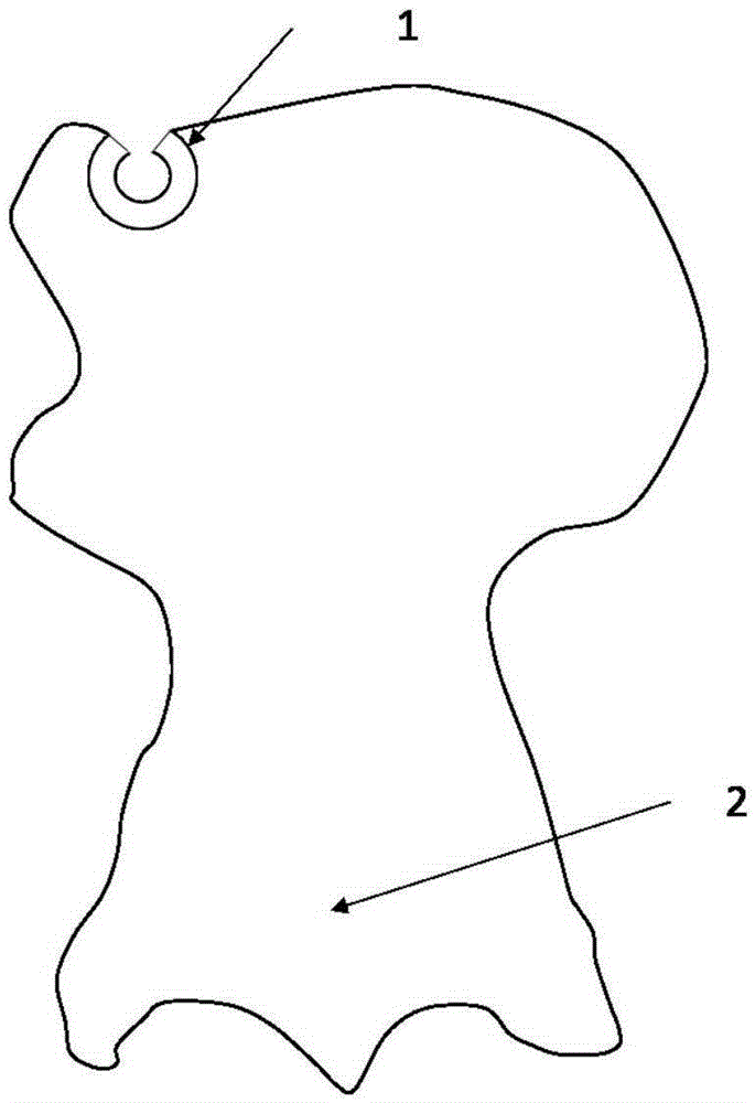
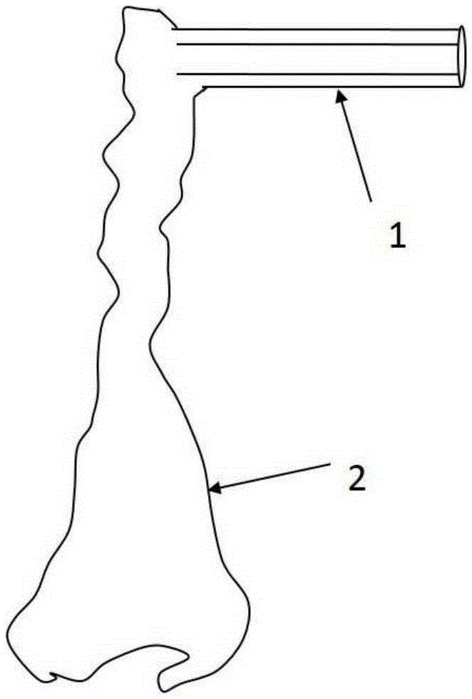
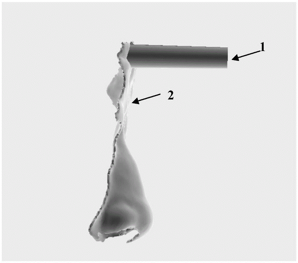
[0005] Aiming at the problems of insufficient existing technology, high cost or poor accuracy, the present invention proposes a 3D printing-based guide bracket for minimally invasive surgery of cerebral hemorrhage, and its preparation method

Method used


Image

  • Guiding stent based on 3D printing and used for cerebral hemorrhage minimally invasive surgery and preparation method thereof
  • Guiding stent based on 3D printing and used for cerebral hemorrhage minimally invasive surgery and preparation method thereof
  • Guiding stent based on 3D printing and used for cerebral hemorrhage minimally invasive surgery and preparation method thereof

Examples


Embodiment 1

[0035] Embodiment 1 A kind of using 3D printing technology to prepare a personalized precision guide bracket for cerebral hemorrhage surgery and its method, the specific method is as follows:

[0036] 1) Thin-layer scanning of the skull of the patient with cerebral hemorrhage is performed by CT, and the DICOM format data file containing the three-dimensional information data of the patient is obtained.

[0037] 2) Store the obtained image data in Dicom format on a CD or DVD disc, import it into Mimics 17.0 medical surgery design software (Materialise, Belgium), select the non-destructive form in the process of importing data, and adjust the operation area face (brow arch) on the working interface , glabella, nose, nasolabial fold, lateral canthus, supraorbital) and hematoma for 3D image modeling.

[0038] 3) Copy the created three-dimensional image model to 3-Matic9.0 (Materialise, Belgium) software, analyze craniofacial and hematoma structures, and design needle entry points ...


PUM

PropertyMeasurementUnit
Thicknessaaaaaaaaaa
Areaaaaaaaaaaa
Lengthaaaaaaaaaa

Abstract

The invention discloses a guiding stent which is obtained by the adoption of 3D printing technology and used for cerebral hemorrhage minimally invasive surgery and preparation method thereof. The preparation method comprises the steps of firstly, scanning the head of a hemorrhage patient so that head image file data can be obtained, then sorting the skull facial profile and target hematoma in need of removal, establishing a three-dimensional model which is closely attached to the skull face of the patient relative to the immobile area profile, designing a hollow non-closed-loop puncture guiding channel stent model in the model, adjusting the direction of a puncture channel, the size of a base and length post-output files of the non-closed-loop puncture channel, conducting 3D printing, and obtaining the accurate guiding stent used for cerebral hemorrhage minimally invasive surgery of the patient. The guiding stent has the advantages that customization of individual characters is achieved, positioning is accurate, infection is avoided, the wound is small, the process is simple, and cost is low.

Description

technical field [0001] The invention relates to the preparation of surgical instruments for cerebral hemorrhage, in particular to a 3D printing-based guide bracket for minimally invasive surgery of cerebral hemorrhage and a preparation method thereof. Background technique [0002] Cerebral hemorrhage has a high morbidity and mortality rate. The healing of cerebral hemorrhage is closely related to the bleeding site, bleeding volume, and rescue time. Cerebral hemorrhage often occurs in areas where important intracranial nuclei are distributed, and these areas are completely different from peripheral bone tissue, craniofacial muscle tissue, and digestive visceral tissue. , they not only pass through important nerve fiber bundles, but also pass through the dense network of blood vessels that dominate the functional area. The operation is extremely difficult and requires extremely high precision in the operation. [0003] With the development of micro-invasive technology, cone h...

Claims


Application Information

Patent Timeline
IPC IPC(8): A61B34/20
Inventor 李泽福李勐杜洪澎李建民刘永良隋德华邵伟王波马立新李珍珠王清波陈正孙其凯耿鑫
Owner 李泽福
Learn More