Fluorescence imaging probe, preparation method and applications thereof

An imaging probe and nano-fluorescence technology, applied in the field of nano-medicine, can solve the problems of indistinguishability and low cure rate of non-small cell cancer patients

Active Publication Date: 2016-03-09
UNIV OF SCI & TECH OF CHINA
View PDF2 Cites 3 Cited by
  • Summary
  • Abstract
  • Description
  • Claims
  • Application Information

AI Technical Summary

Problems solved by technology

However, at present, the two types of cells cannot be accurately distinguished by clinicopathological characteristics.
Physicians usually make judg...

Method used


Image

  • Fluorescence imaging probe, preparation method and applications thereof
  • Fluorescence imaging probe, preparation method and applications thereof
  • Fluorescence imaging probe, preparation method and applications thereof

Examples


preparation example Construction

[0053] In some specific embodiments of the present invention, the preparation method of nanometer fluorescent imaging probe comprises the following steps:

[0054] Prepare uniform manganese cobalt cyanate nanoparticles: take 0.15mmol manganese acetate (Mn(CH3COO)2.4H2O) (0.036765g), 0.6gPVP is dispersed in the mixed solution of ethanol and distilled water (15mL ethanol, 5mL distilled water), 0.08mmol Potassium cobaltcyanide (K3[Co(CN)6)]) (0.0265864g) was dispersed in 6mL of distilled water, and then the solution was added dropwise to the above mixed solution, and stirred for 10min to obtain manganese cobaltcyanate nanoparticles.

[0055] Preparation of uniform cadmium-doped manganese cobalt cyanate nanoparticles: 10 mL of distilled aqueous solution containing 0.02 mmol cadmium chloride CdCl (0.0036 g) was added to the prepared manganese cobalt cyanate nanoparticles, and continued to stir for 3 h, then the solution Let stand for 24 hours, and finally centrifuge and wash to obt...

Embodiment 1

[0061] Take 0.15mmol manganese acetate (Mn(CH 3 COO) 2 4H 2 O) (0.036765g), 0.6gPVP is dispersed in the mixed solution of ethanol and distilled water (15mL ethanol, 5mL distilled water), 0.08mmol potassium cobalt cyanide (K 3 [Co(CN) 6 )]) (0.0265864g) was dispersed in 6mL of distilled water, and then the solution was added dropwise to the above mixed solution, and stirred for 10min to obtain uniform manganese cobalt cyanate nanoparticles.

Embodiment 2

[0063] 10mL containing 0.02mmol cadmium chloride CdCl 2 The distilled aqueous solution of (0.0036g) joins in the manganese cobalt cyanate solution that makes by embodiment 1, and continues to stir 3h, then solution is left standstill 24h, finally centrifugal washing, promptly obtains uniform cadmium doped manganese cobalt cyanate Nanoparticles (7% content of cadmium ions in the nanofluorescence imaging probe).

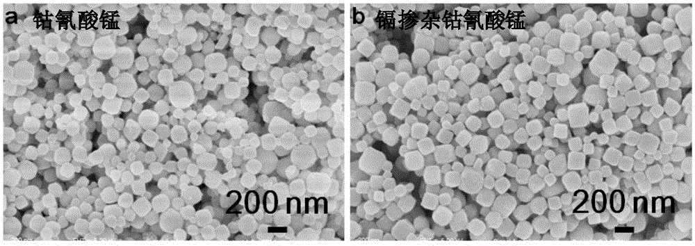
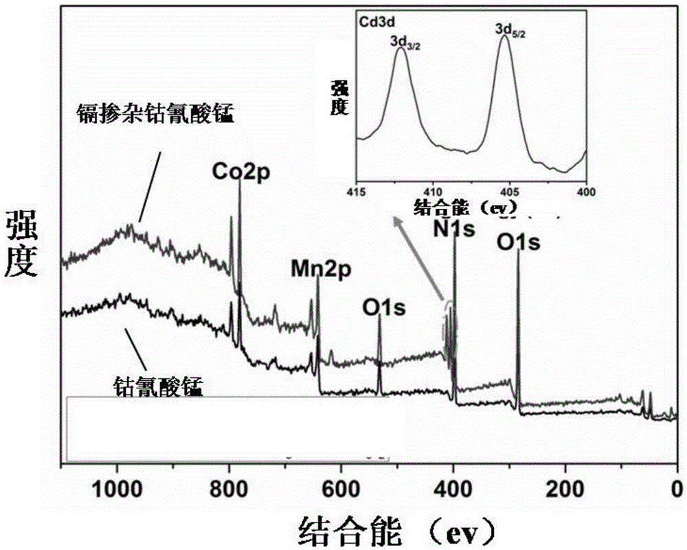
[0064] According to the scanning electron microscope image ( figure 1 ) It can be seen that the product has good dispersibility. After cadmium ions are doped into the manganese cobalt cyanate nanoparticles, the structure of the original nanoparticles is not affected, and the X-ray photoelectron spectroscopy analysis diagram ( figure 2 ) shows that after doping, the characteristic peak of cadmium element appears in the product, which proves that cadmium ions are indeed doped into manganese cobalt cyanate nanoparticles.


PUM


Abstract

The invention relates to the field of nano medicine, and especially relates to a fluorescence imaging probe, a preparation method and applications thereof. The nano fluorescence imaging probe comprises a material having a metal-organic skeleton structure and heavy metal ions. The probe can enter lung cancer cells through endocytosis. The nano fluorescence imaging probe can stimulate different lung cancer cells to generate different contents of metallothionein, then the probe can combine with metallothionein, then the probe and metallothionein are discharged from the cells, and the amount of residual nano fluorescence imaging probe in cells will determine the intensity of cell fluorescence images, so according to the difference of intensity of cell fluorescence images, the lung squamous cell carcinoma and lung adenocarcinoma can be precisely differentiated.

Description

technical field [0001] The invention relates to the field of nanomedicine, in particular to a fluorescent imaging probe and its preparation method and application. Background technique [0002] Lung cancer is a malignant tumor disease with a relatively high incidence rate, which seriously threatens human life. According to tumor histological characteristics, lung cancer can be divided into non-small cell carcinoma and small cell carcinoma. These two types of lung cancer can be accurately distinguished based on their clinicopathological features. Non-small cell carcinoma can be further divided into lung squamous cell carcinoma and lung adenocarcinoma (Lung Cancer, volume 3, pages 326-327). Accurately distinguishing these two types of non-small cell carcinoma is very important for evaluating the treatment effect of patients, formulating a reasonable treatment plan, and choosing a reasonable treatment drug. However, these two types of cells cannot be accurately distinguished...

Claims


Application Information

Patent Timeline
IPC IPC(8): G01N21/64
Inventor 陈乾旺陈健
Owner UNIV OF SCI & TECH OF CHINA
Why Eureka
Try Eureka