Biochemical marker for CVD risk evaluation

A bioassay, C-terminal technology, applied in the field of biochemical markers for CVD risk assessment

Active Publication Date: 2016-04-20
NORDIC BIOSCI
View PDF3 Cites 1 Cited by
  • Summary
  • Abstract
  • Description
  • Claims
  • Application Information

AI Technical Summary

Problems solved by technology

However, in some cases, a more invasive approach,

Method used


Image

  • Biochemical marker for CVD risk evaluation
  • Biochemical marker for CVD risk evaluation
  • Biochemical marker for CVD risk evaluation

Examples


Embodiment 1

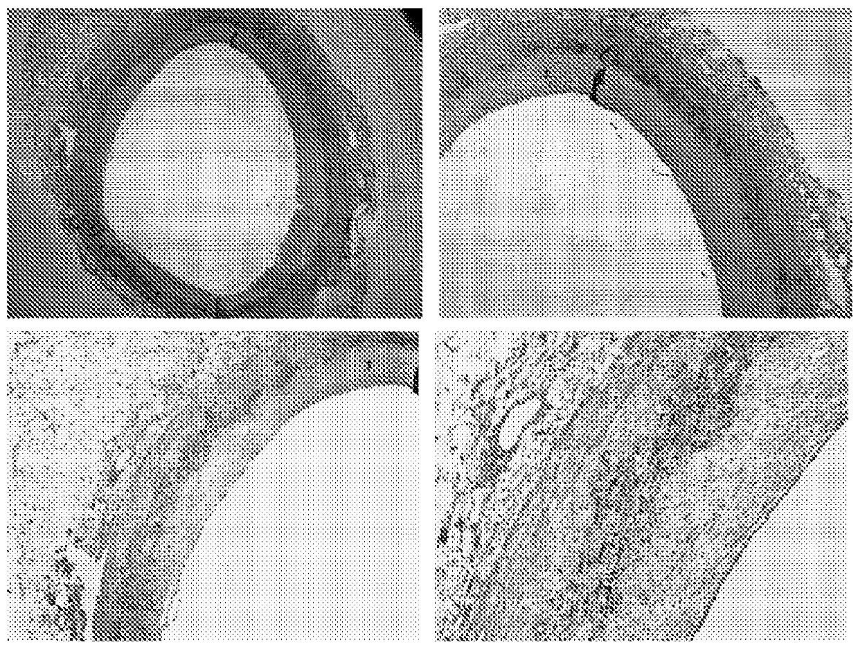
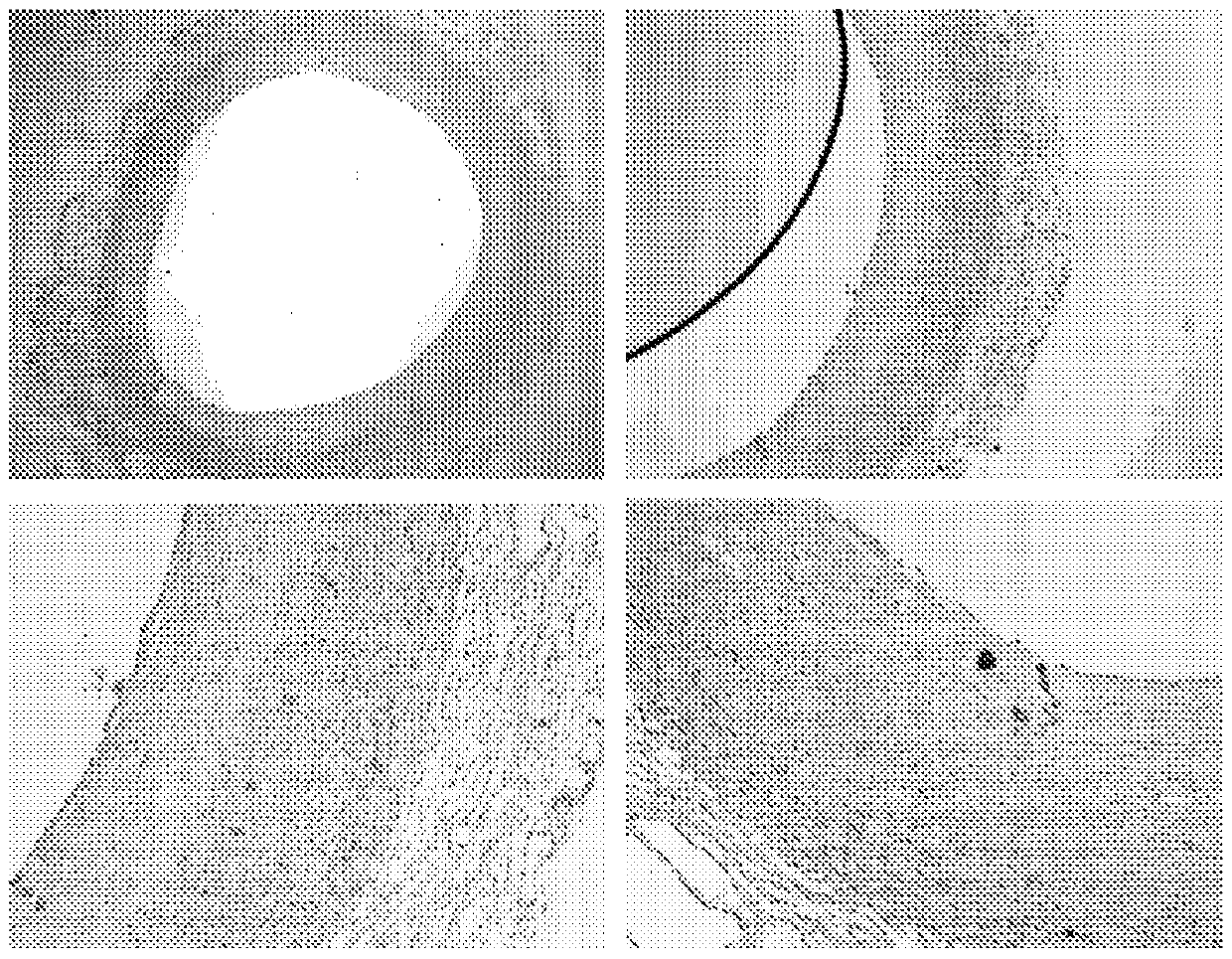
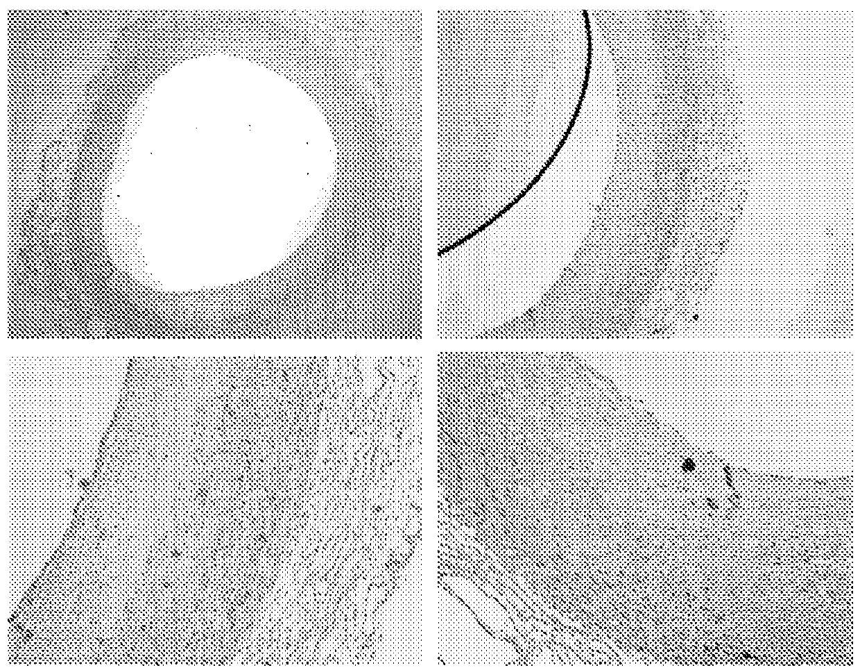
[0265] To analyze the localization of proteoglycans and proteases, we performed immunohistochemical staining on human arterial samples from left anterior descending coronary arteries (LAD). In the following, it is demonstrated that the cathepsin K protease colocalizes with biglycan.

[0266] Such as figure 1 with 2 Immunohistochemical staining shown in shows colocalization of biglycan with cathepsin K. This may indicate that biglycan is a preferred substrate for cathepsin K. The same immunohistochemical staining was performed on samples of aorta in which atherosclerotic plaques formed and consequently resulted in macrophage foam cell infiltrates and calcifications replacing normal aortic structures. The results of these immunostainings are summarized in image 3 with 4 middle.

[0267] Immunohistochemical staining of biglycan and cathepsin K revealed co-localization in developing atherosclerotic lesions. Combining these results led to the hypothesis that a specific cle...

Embodiment 2

[0269] Biglycan degradation for assessment of degraded fragments. Biglycan from bovine articular cartilage (B8041-Sigma-Aldrich) was cleaved by the following proteases: MMP2, MMP3, MMP8, Cathepsin K, Cathepsin S, Cathepsin B, and Cathepsin L. The proteoglycan fragments produced by the enzymatic cleavage of the above proteases were treated at 10% Separation on a Bis-Tris gel followed by silver staining by the "SilverExpress" silver staining kit (Invitrogen Cat# LC6100, Lot# 341099). Separation of proteolytically produced biglycan and silver staining results in Figure 5 displayed in .

Embodiment 3

[0271] Mice were immunized with peptides from type III collagen conjugated to ovalbumin. Serum was screened for reactivity with biotin-conjugated screened peptide sequences. Monoclonal antibody-secreting clones are generated and screened using the selection sequence. Clones were examined for lack of reactivity with an extended form of the target peptide that continues an adjacent sequence in type III collagen (reverse selection peptide) and for lack of reactivity with a nonsense peptide. All target sequence positive clones did not react with extended or nonsense sequences.

[0272] The target sequence, immunogen, screening sequence and counter-selection sequence are as follows:

[0273]


PUM


Abstract

The invention relates to a biochemical marker for CVD risk evaluation and a method of bioassay for the quantification of peptide fragments comprising a neo-epitope formed by cleavage of a protein of an atherosclerotic plaque such as lumican, versican, perlecan, decorin, biglycan, collagen type III, CRP, ApoE, or elastin, by a proteinase, said comprises contacting a sample such as urine or serum with an antibody reactive with the neo-epitope and determining the level of binding of said immunological binding partner to peptide fragments in said sample. The assay is predictive of risk of cardiovascular disease events.

Description

[0001] This application is a divisional application of Chinese patent application CN200880123912.0, and the original application is an application that entered the Chinese national phase on July 2, 2010 with the international application number PCT / EP2008 / 064946. technical field [0002] The present invention relates to assays for the detection of biochemical markers valuable for diagnostic purposes of cardiovascular disease and prognosis of disease progression, including biochemical markers indicative of the risk of cardiovascular events resulting from the development of atherosclerosis and plaque instability things. Background technique [0003] Cardiovascular disease (CVD) is the leading cause of disease and death worldwide. Currently, there is no efficient and non-invasive diagnostic method that allows diagnosis and classification of patients into different risk groups, as well as allowing diagnosis of low-risk patients. Diagnostic and prognostic tools primarily consist...

Claims


Application Information

Patent Timeline
IPC IPC(8): G01N33/68
CPCG01N33/68C07K14/775G01N33/542G01N2800/32C12Q1/37C07K14/77C07K14/78G01N33/577G01N33/6878G01N33/6893G01N2800/50G01N2800/52
Inventor 莫滕·卡尔斯达尔佩尔·奎斯特娜塔莎·巴拉斯丘克
Owner NORDIC BIOSCI
Why Eureka
Try Eureka