Method for three-dimensional registration and brain tissue extraction of individual human brain multimodality medical images

A medical imaging and three-dimensional registration technology, which is used in medical science, instruments for radiological diagnosis, diagnosis, etc. It can solve the difference of electrode position, cannot adjust the color setting of PET imaging, and can only perform single image registration in stages, etc. question

Inactive Publication Date: 2016-08-03
王雪原 +1
View PDF11 Cites 31 Cited by
  • Summary
  • Abstract
  • Description
  • Claims
  • Application Information

AI Technical Summary

Problems solved by technology

[0003] For patients after implantation of intracranial electrodes, in order to show the relationship between the exposed cerebral cortex, buried intracranial electrodes and the overall shape of the brain, many methods have been developed, listed as follows: The first method is to Digital photographs are fused with magnetic resonance imaging (MRI) images, but the reconstructed cortex has poor morphology and lacks blood vessels as positioning marks. Since some electrodes are located outside the bone window, it is impossible to connect them with the cortical surface even if intraoperative photographs are taken. Carry out position-relationship correspondence, which is detrimental both for electrical stimulation mapping of cortical functions and for preservation of important functions during surgery
The second method is to use the cranial X-ray after intracranial electrode implantation and preoperative MRI for registration, which can show the position of the intracranial electrode on the cortex, but the electrodes at the skull base and midline cannot be well displayed
The fourth method is to use the self-edited Matalab program to sample intraoperative photos, and draw the brain surface curve to obtain the intracranial electrode position, but there are also differences with the actual electrode position, and the electrodes that are blocked outside the bone window are still unable to be located
The

Method used


Image

  • Method for three-dimensional registration and brain tissue extraction of individual human brain multimodality medical images
  • Method for three-dimensional registration and brain tissue extraction of individual human brain multimodality medical images
  • Method for three-dimensional registration and brain tissue extraction of individual human brain multimodality medical images

Examples


Embodiment Construction

[0038] The present invention will be described in detail below in conjunction with the accompanying drawings and embodiments.

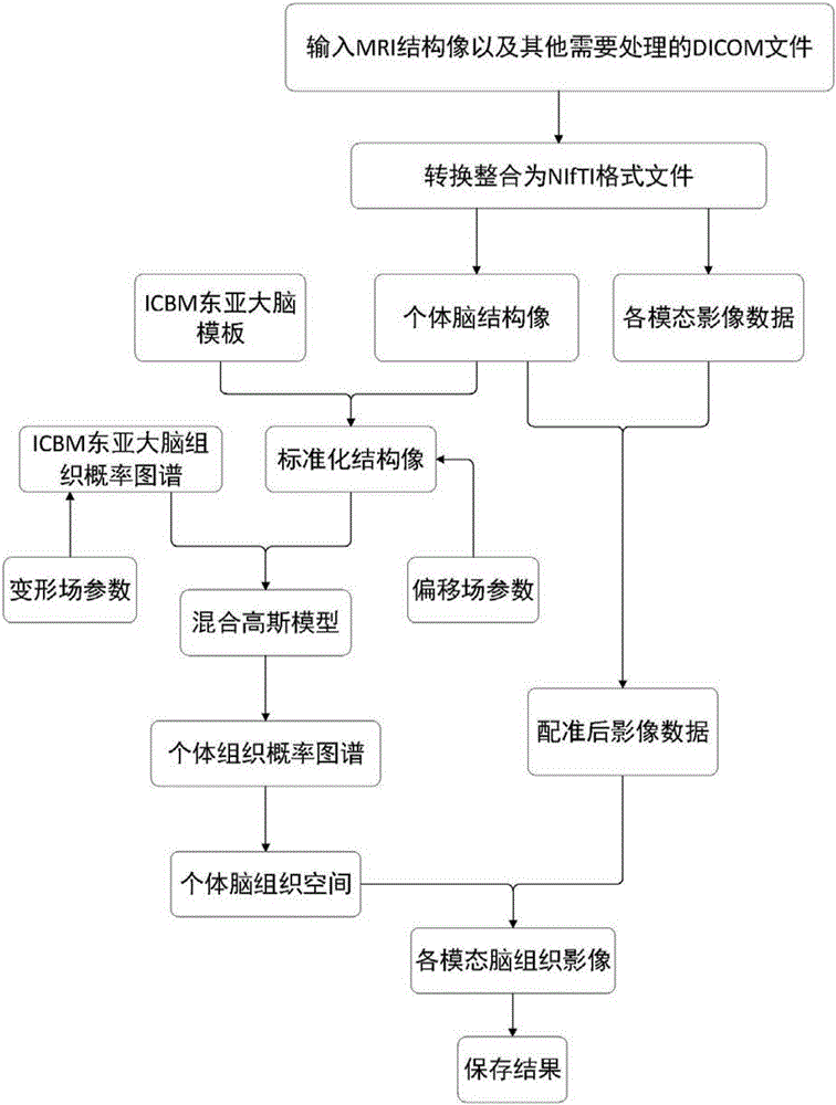
[0039] Such as figure 1 As shown, the present invention provides a method for three-dimensional registration and brain tissue extraction of individualized multimodal medical images of the human brain, which includes the following steps:

[0040] (1) Read DICOM medical image data, and convert DICOM format data into NIfTI format;

[0041] (1.1) Convert the nuclear magnetic resonance structure image (3TT1-MPR) DICOM format data into NIfTI format and save it;

[0042] (1.2) Each modality (such as CT, MRA, MRV, T2-flair, MEG, 18 FDG-PET metabolic imaging, etc.) DICOM format image data is converted to NIfTI format file and saved;

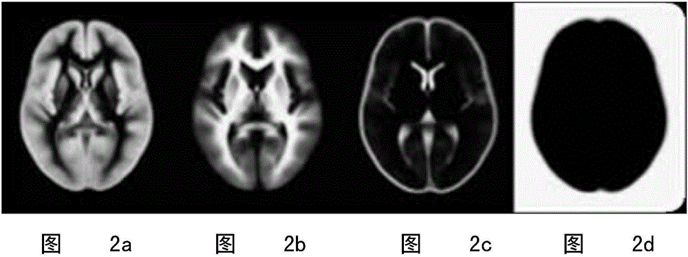
[0043] (2) Layering the NMR structural image: Using the ICBM (internationalconsortiumforbrainmapping, Human Brain Mapping Alliance) East Asian brain structure template and East Asian brain tissue probability atlas, a mixed Gau...


PUM


Abstract

The invention relates to a method for three-dimensional registration and brain tissue extraction of individual human brain multimodality medical images. The method comprises the following steps: reading DICOM medical image data, and converting DICOM format data into NIfTI format data; layering images adopting a nuclear magnetic resonance structure; establishing a mixture gaussian model through an east Asia brain structure template and an east Asia brain tissue probability graph of ICBM, and dividing nuclear magnetic data into a grey matter part, a white matter part and a cerebrospinal fluid part; enabling the registration methods of other modality data and nuclear magnetic structure images to be the same; according to a layering and registration result, removing a skull and other parts outside the skull of each modality, and reserving the structure of parts inside the skull; adding weighting of three tissue probability graphs of the grey matter, the white matter and the cerebrospinal fluid obtained through layering of structure images so as to obtain a brain tissue probability graph, and performing Gaussian kernel smoothness; setting a threshold, applying the threshold to each modality image data after registration and resampling, and removing the cranium and parts outside the cranium; outputting a save result in an NIfTI format. According to the method disclosed by the invention, the registration of various structure images and multiplanar reconstruction images of a tested person can be completed at the same time.

Description

technical field [0001] The invention relates to a three-dimensional image registration and brain tissue extraction method in the medical field, in particular to a three-dimensional registration and brain tissue extraction method of individualized multimodal medical images of the human brain. Background technique [0002] Epilepsy surgery involves localization of epileptogenic focus, cerebral cortex function localization and intracranial electrode localization. The characteristic EEG signals of the seizure period and the cortical electrical stimulation results collected by the intracranial electrodes need to be mapped to the corresponding structures of the cerebral cortex, which is obviously helpful for the localization of the epileptogenic focus and the formulation of the surgical resection range. The solution of these problems is inseparable from the development of medical imaging and computer technology. [0003] For patients after implantation of intracranial electrodes,...

Claims


Application Information

Patent Timeline
IPC IPC(8): A61B6/00
CPCA61B6/5247
Inventor 王雪原温建斌李小俚遇涛张国君李勇杰
Owner 王雪原
Why Eureka
Try Eureka