Dual-mode optical imaging probe with tumor double-targeting function and preparation and application thereof
An optical imaging, dual-targeting technology, applied in the field of SERS-fluorescence dual-mode optical imaging probes, can solve problems such as the limitations of fluorescence multiplex imaging, and achieve the effect of enhancing target recognition and efficiency, high sensitivity, and improving endocytosis
- Summary
- Abstract
- Description
- Claims
- Application Information
AI Technical Summary
Problems solved by technology
Method used
Image
Examples
Embodiment 1
[0045] To prepare a dual-mode probe with enhanced targeting effect on cervical cancer cells (HeLa), the preparation method is as follows:
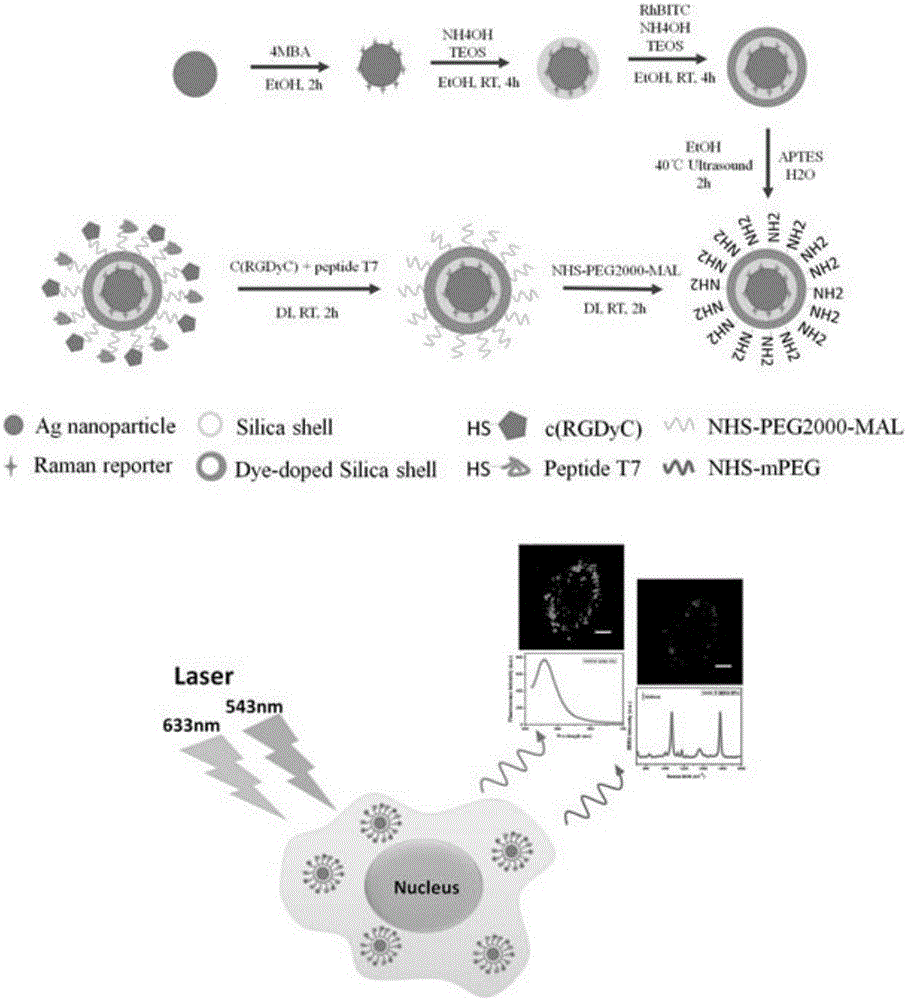
[0046] 1) The experiment uses the seed growth method to prepare a uniform spherical silver nanoparticle solution. Under vigorous stirring, 5 mL of 1% sodium citrate aqueous solution was added to boiling 245 mL of silver nitrate solution (containing 90 mg of silver nitrate), and stirring was continued for one hour to obtain a silver glue solution. Take 10mL of silver gel solution and disperse it in alcohol solution by centrifugation, add 10uL of 10mM Raman reporter 4MBA, mix and stir for 1-4 hours, observe whether the color of the solution changes to prevent aggregation;
[0047] 2) Add 200μL of ammonia water and 5μL of tetraethoxysilane solution to the solution of step 1, and stir at room temperature for more than 4 hours, and observe that the solution turns into khaki;
[0048] 3) Add 200μL of ammonia, 5μL of 1mg / mL 3-aminopropyltriethoxysilane...
Embodiment 2
[0053] Furthermore, the dual-targeting dual-mode fluorescent probe is used in dual-mode imaging of cervical cancer cells HeLa, the specific method is as follows:
[0054] 1) Inoculate cervical cancer cells in a petri dish, add MEM cell culture medium containing 10% fetal bovine serum and 1% penicillin-streptomycin mixture, and place it in an incubator at 37°C and 5% CO2 for 24 hour. After that, add the probe at a 4:1 ratio of culture solution to probe, mix well and place it in the incubator for two hours. After the particles are internalized by the cells, the culture medium is removed, the cells are washed 3 times with PBS, fixed with 4% paraformaldehyde for 10 minutes, and washed twice with PBS for later use.
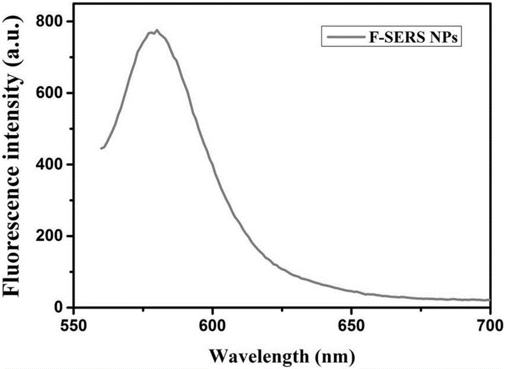
[0055] 2) Add a small amount of buffer to the fixed cells obtained in step 1, adjust the focal plane to focus, select a cell area, excite with 543nm laser, 560nm-600nm as the receiving wavelength range, imaging to obtain cell fluorescence images, such as Figure 5 Shown. ...
Embodiment 3
[0058] Furthermore, in order to show that the targeting ability of the dual-targeting probe is better than that of the traditional single-targeting probe, the following experiments were performed to verify its targeting ability:
[0059] 1) Prepare a single-targeting probe according to the steps in Example 1. The only difference is that in the last step, only one polypeptide of the same concentration as the double-targeting probe is added to the solution. Here, cys-HAIYPRH is selected as an example.
[0060] 2) Inoculate cervical cancer cells in two culture dishes at the same concentration, add MEM cell culture medium containing 10% fetal bovine serum and 1% penicillin-streptomycin mixture, and place in an incubator at 37°C, 5% CO 2 Cultivate for 24 hours in the environment. After that, the two probes were added with a 4:1 ratio of culture solution to probe, mixed evenly and then placed in the incubator for two hours. After the particles are internalized by the cells, the culture m...
PUM
 Abstract
Description
Claims
Application Information
 - R&D
- Intellectual Property
- Life Sciences
- Materials
- Tech Scout
- Unparalleled Data Quality
- Higher Quality Content
- 60% Fewer Hallucinations
Browse by: Latest US Patents, China's latest patents, Technical Efficacy Thesaurus, Application Domain, Technology Topic, Popular Technical Reports.
© 2025 PatSnap. All rights reserved.Legal|Privacy policy|Modern Slavery Act Transparency Statement|Sitemap|About US| Contact US: help@patsnap.com


