Isolation and culture method for primary mice or rat cardiac muscle cells

A technology for the separation and cultivation of cardiomyocytes, applied in the field of cell culture, can solve the problems of increased bacterial, cell, and fungal contamination, difficulty in cardiac perfusion operations, insufficient clear blood contamination, etc., to achieve great promotional significance, avoid contamination, and evenly digest thorough effect

Inactive Publication Date: 2016-08-31
王晓冰 +1
View PDF5 Cites 10 Cited by
  • Summary
  • Abstract
  • Description
  • Claims
  • Application Information

AI Technical Summary

Problems solved by technology

And with the passage of cells, the number of cardiomyocytes will decrease, and the proportion of myocardial fibrosis will increase, which will seriously affect the experimental results.
[0005] Secondly, due to the particularity of the heart collection process, even external flushing plus the heart’s own rhythmic contraction to discharge most of the blood in the heart is still not enough to clear the blood contamination, so when the myocardial cell tissue pieces are removed, a small amount of blood is often mixed. This part of the blood greatly increases the possibility of contamination by various bacteria, cells, and fungi in the subsequent cell culture process.
In order to completely overcome the influence of this part of blood contamination, those skilled in the art often use the method of cardiac perfusion. However, the cardiac perfusion operation of mice / rats is difficult and time-consuming, and is not suitable for application.

Method used


Image

  • Isolation and culture method for primary mice or rat cardiac muscle cells
  • Isolation and culture method for primary mice or rat cardiac muscle cells

Examples


Embodiment 1

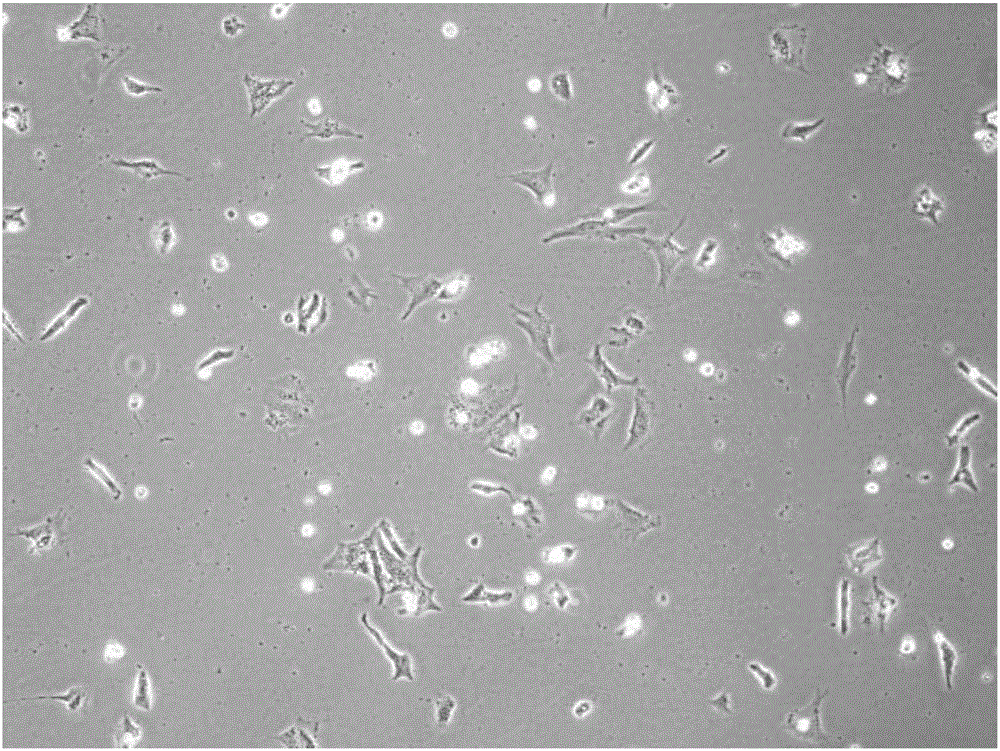
[0039] In this embodiment, SD rats are adopted, and the separation and culture method of the present invention is used to separate cardiomyocytes, and the specific steps are as follows:

[0040] 1. Rat Heart Harvesting

[0041] Firstly, newborn rats were sacrificed (cervical dislocation), soaked in alcohol for 5 minutes, and under a dissecting microscope, the chest cavity was opened to fully expose the heart. Use tweezers to clamp the root of the heart, the blood vessels near the atrium, and cut out the complete heart. Try not to clamp the tissue of the ventricle, because the cardiomyocytes are very sensitive to the clamp, which will easily lead to the death of the cardiomyocytes at the clamped site. The pericardium and other accompanying connective tissue were cleaned and irrigated with D-hanks solution containing double antibody and amphotericin B.

[0042] 2. Take cardiomyocytes

[0043] Under a dissecting microscope, a small biopsy needle is inserted into the wall of the...


PUM


Abstract

The invention discloses an isolation and culture method for primary mice or rat cardiac muscle cells. The isolation and culture method employs specially-prepared compound enzyme for long-term digestion, so the obtained cells are large in gross quantity and high in both isolation degree and motility rate; due to different acquisition manners, pollution and influence of residual blood in a heart are eradicated and the probability of contamination of the acquired cardiac muscle cells by bacteria, fungi and other cells is low; and the method can realize subculturing, so a good cell culture scheme is provided for related research based on culture of cardiac muscles.

Description

technical field [0001] The invention relates to the technical field of cell culture, in particular to a method for isolating and culturing primary mouse or rat cardiomyocytes. Background technique [0002] Cardiomyocytes are the most energy-rich cells in the body. They are highly oxygenated cells and have a large number of mitochondria. Cardiomyocytes make up 75% of the volume of the heart, but only make up one-third of the total number of cells in the heart. Although differentiated cardiomyocytes are difficult to proliferate, however, hypertrophy can occur in the heart upon stimulation by alpha-1 adrenaline through the Ras / MEK pathway. The membrane of all cardiomyocytes can spontaneously produce rhythmic depolarization and repolarization. The contraction of cardiomyocytes is myogenic and does not depend on nerve stimulation. There is a complex network of signaling systems in cardiomyocytes that regulate the rhythmic contraction and contraction of the heart. The loss of ...

Claims


Application Information

Patent Timeline
Patent Type & Authority Applications(China)
IPC IPC(8): C12N5/077
CPCC12N5/0657C12N2509/10
Inventor 王晓冰杨国峰
Owner 王晓冰
Why Eureka
Try Eureka