3D-priniting extracorporeal-assisted biopsy/positioning device and manufacture method thereof

A 3D printing and positioning device technology, applied in computer-aided planning/modeling, inoculation and ovulation diagnosis, medical science, etc., can solve problems such as aggravating the mental burden of patients, limited movable range of puncture needles, and radiation effects of patients. Risk of complications, avoidance of inaccessible nodules, effect of improved puncture accuracy

Active Publication Date: 2016-11-09
上海揽慧医疗科技有限公司
View PDF7 Cites 8 Cited by
  • Summary
  • Abstract
  • Description
  • Claims
  • Application Information

AI Technical Summary

Problems solved by technology

[0007] However, the existing preoperative positioning technology has the following problems: 1. The mass cannot be touched by hand during the operation: confirming the position of the mass by touching during the operation is theoretically the most direct and accurate method, but in many cases Due to various reasons such as the texture of the lung mass is tough, the volume of the mass is too small, the position of the mass is deep, and the doctor is inexperienced, etc., the manual palpation may not be palpable or the touch is inaccurate, so that the lung mass cannot be completely removed; 2. Radiation radiation Influence: Preoperative CT is guided by hook-wire positioning needle or micro-coil microcoil for puncture positioning. It is necessary to undergo multiple CTs in a short period of time to determine the positioning position and cause radiation impact on patients; 3. Staffing

Method used


Image

  • 3D-priniting extracorporeal-assisted biopsy/positioning device and manufacture method thereof
  • 3D-priniting extracorporeal-assisted biopsy/positioning device and manufacture method thereof
  • 3D-priniting extracorporeal-assisted biopsy/positioning device and manufacture method thereof

Examples


preparation example Construction

[0043] The present invention also provides a preparation method of a 3D printed in vitro assisted biopsy / positioning device, comprising the following steps:

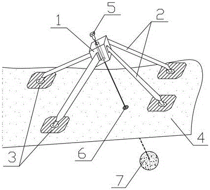
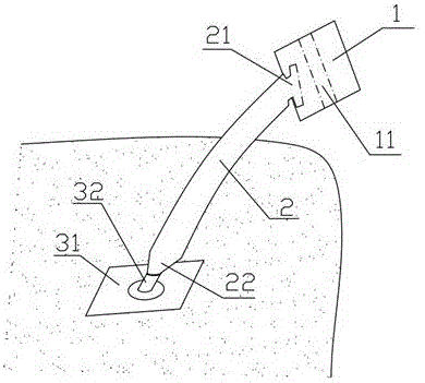
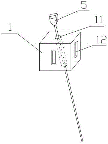
[0044] (1) Based on the pre-processed positioning slice image with the lung mass, use modeling software to reconstruct the model of the patient's chest cavity and lungs with mass, and determine the projection of the lung mass on the body surface on the chest cavity and lung models Location;

[0045] (2) According to the pre-calculated needle insertion angle and depth, use the modeling software again to build a fitting umbrella biopsy / positioning model including the central positioning module model, antennae model and fixed tripod model on the above chest and lung models ;

[0046] (3) Import the center positioning module, antennae model and fixed tripod model into the printer and perform 3D printing to obtain the positioning module model, antennae and fixed tripod;

[0047] (4) Assemble the 3D-printed positioning modul...


PUM


Abstract

The invention discloses a 3D-priniting extracorporeal-assisted biopsy/positioning device. The biopsy/positioning device is formed by 3D printing. The 3D-priniting extracorporeal-assisted biopsy/positioning device comprises a central positioning module, in which a needle-feeding hole duct used for feeding a puncture needle is arranged. The surface of the central positioning module is provided with a first clamping slot. The 3D-priniting extracorporeal-assisted biopsy/positioning device further comprises an antenna with one end having a first clamping pin in snap joint with the first clamping slot and with the other end having a second clamping pin, a fixing foot stand comprising a base and support feet arranged on the base. The support feet and a plane where the base is located are arranged at certain angle. One end, away from the base, of the support feet is provided with a second clamping slot. The second clamping slot is in snap joint with the second clamping pin. The 3D-priniting extracorporeal-assisted biopsy/positioning device is broad in application scope, short in operation time and helps relive pains of a patient and achieves prices positioning and puncture biopsy of preoperative lumps.

Description

technical field [0001] The invention relates to a positioning device for a pulmonary mass in laparoscopic surgery, in particular to a 3D printed in vitro auxiliary biopsy / positioning device and a preparation method thereof. Background technique [0002] In clinical work, clinical problems that require the use of needle biopsy techniques or preoperative positioning techniques are often encountered. Needle biopsy can clarify the histological type of the lesion, determine the pathological diagnosis, and guide clinical treatment. The preoperative positioning technique of the mass is very important to determine the scope of resection during operation. [0003] The above clinical issues include lung tumor biopsy and preoperative localization, thyroid biopsy, breast biopsy, and liver biopsy lesions. [0004] The existing technologies of preoperative positioning and needle biopsy will be introduced respectively as follows: [0005] 1. Preoperative positioning: Taking lung tumors ...

Claims


Application Information

Patent Timeline
IPC IPC(8): A61B17/34A61B34/10A61B10/04
Inventor 范子文西尔买买提·卡德尔李泽遥李牧王龙顾亚伟陈昶
Owner 上海揽慧医疗科技有限公司
Why Eureka
Try Eureka