Targeted-imaging agent used for detecting glioma and preparation method and application of targeted-imaging agent

A targeted imaging agent and glioma technology, applied in the field of imaging medicine, can solve the problems of low resolution of SPECT imaging images, inability to diagnose and grade gliomas, difficulty in displaying early small tumors, etc., and achieve high resolution High-resolution imaging, high-resolution imaging, and high-sensitivity effects

Inactive Publication Date: 2017-01-25
THE FIRST AFFILIATED HOSPITAL OF SUN YAT SEN UNIV
View PDF1 Cites 3 Cited by
  • Summary
  • Abstract
  • Description
  • Claims
  • Application Information

AI Technical Summary

Problems solved by technology

[0003] The problem with cranial magnetic resonance examination is that it cannot diagnose and grade glioma well
Specifically: For the convenience of image analysis of glioma, a weighted image appears. A weighted image is an image that determines the grayscale of a brain MRI image through a specific imaging parameter. Cranial MRI The examination shows the glioma in the brain through the weighted image, but the weighted image can only reflect the degree of damage to the blood-brain barrier of the brain glioma, but cannot accurately show the boundary of the brain glioma
[0004] There are two problems with SPECT imaging. One is that SPECT imaging uses planar imaging, and there is a phenomenon of radioactive superimposition. The second is that the image resolution of SPECT imaging is low, and it is difficult to display early small tumors (<2.5cm)

Method used


Image

  • Targeted-imaging agent used for detecting glioma and preparation method and application of targeted-imaging agent
  • Targeted-imaging agent used for detecting glioma and preparation method and application of targeted-imaging agent
  • Targeted-imaging agent used for detecting glioma and preparation method and application of targeted-imaging agent

Examples


preparation example Construction

[0039] Main experimental instruments and experimental materials in the preparation method of the imaging agent of the embodiment of the present invention include:

[0040] The SEP-PAK light QMA column is a solid phase extraction column produced by Waters Corporation of the United States;

[0041] Amino polyether, the English name is Kryptofix222 (abbreviated as K 222 ), produced by ABX Company of Germany;

[0042] HLB is a hydrophilic-lipophilic balanced adsorbent, which is a modified styrene divinylbenzene copolymer;

[0043] 0.01M PBS (PH=7.4), produced by Guangzhou Zhanchen Biotechnology Co., Ltd.;

[0044] Anhydrous acetonitrile (MeCN), produced by U.S. Sigma-Aldrich company;

[0045] Chlorotoxin (Chlorotoxin), produced by Japan Peptide Institute, Inc.;

[0046] Sep-Pak C18 column, produced by American Waters Company;

[0047] Fluorine multi-functional chemical synthesis module (PET-MF-2V-IT-I), produced by Beijing Petrochemical Co., Ltd.;

[0048] Inveon Micro PET-C...

Embodiment 1

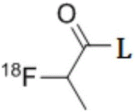
[0060] The targeted imaging agent used to detect glioma in this embodiment is 18 F-fluorobenzoic acid-chlorotoxin, English as 18 F-FP-Chlorotoxin, its structural formula is:

[0061]

[0062] Wherein, L is the amino acid sequence shown in SEQ ID NO:1.

[0063] The preparation method of the targeted imaging agent for detecting glioma in this embodiment comprises the following steps:

[0064] (1) Synthesis of 18F-N-succinimide 4-[18F]fluorobenzoic acid;

[0065] (2) 18 F-N-succinimide 4-[ 18 F] coupling reaction of fluorobenzoic acid and chlorotoxin;

[0066] Wherein, step (1) comprises the following steps in order:

[0067] (A) Preparation 18 f - : passed by the cyclotron 18 O(p,n) 18 F nuclear reaction to generate 18 F-F - , under He gas transmission, through the SEP-PAK light QMA column placed in the radioactive detector, 18 f - Trapped on the SEP-PAK light QMA column;

[0068] (B) Preparation [K / K 222 ] 18 f - : Under the action of vacuum pump, use 1mL o...

Embodiment 2

[0076] The targeted imaging agent used to detect glioma in this example is the same as that in Example 1.

[0077] The preparation method of the targeted imaging agent for detecting glioma in this embodiment comprises the following steps:

[0078] (1) Synthesis of 18F-N-succinimide 4-[18F]fluorobenzoic acid;

[0079] (2) 18 F-N-succinimide 4-[ 18 F] coupling reaction of fluorobenzoic acid and chlorotoxin;

[0080] Wherein, step (1) comprises the following steps in order:

[0081] (A) Preparation 18 f - : passed by the cyclotron 18 O(p,n) 18 F nuclear reaction to generate 18 F-F -, under He gas transmission, through the SEP-PAK light QMA column placed in the radioactive detector, 18 f - Trapped on the SEP-PAK light QMA column;

[0082] (B) Preparation [K / K 222 ] 18 f - : Under the action of vacuum pump, use 1mL of K 222 (Amino polyether) solution will 18 f - Rinse to form a mixed solution in the first reaction bottle, pass nitrogen gas (100ml / min) into the fir...


PUM


Abstract

The invention relates to the technical field of medical imaging technology, in particular to a targeted-imaging agent used for detecting glioma and preparation method and application of the targeted-imaging agent. The targeted-imaging agent refering to <18>F-fluorobenzoic acid-chlorotoxin has the advantages that boundaries of the glioma can be accurately defined; high sensitivity is achieved due to the fact that the chlorotoxin marked with <18>F can be subjected to imaging detection via PET (positron emission tomography) and focus 6mm in size can be detected with the PET which is much higher than SPECT (single photon emission computed tomography) in image resolution; a brand new direction is provided for diagnosis the glioma, and important clinical value in aspects of early diagnosis of the glioma, boundary define of the glioma, therapeutic evaluation, targeted drug delivery and the like is achieved; the targeted-imaging agent can be applied to the PET, the boundaries of the glioma can be accurately defined, high image resolution can be realized, the focus 6mm in size can be detected, and the PET is much higher than the SPECT in the image resolution in the prior art.

Description

technical field [0001] The invention relates to the technical field of imaging medicine, in particular to a targeted imaging agent for detecting glioma and its preparation method and application. Background technique [0002] Glioma is a tumor derived from the neuroectoderm, accounting for 40-50% of brain tumors, and is the most common intracranial malignant tumor. The most commonly used methods for non-invasive diagnosis of glioma in clinical practice are cranial magnetic resonance examination and SPECT imaging. [0003] The problem with cranial MRI is that it cannot diagnose and grade glioma well. Specifically: For the convenience of image analysis of glioma, a weighted image appears. A weighted image is an image that determines the grayscale of a brain MRI image through a specific imaging parameter. Cranial MRI The examination shows the glioma in the brain through the weighted image, but the weighted image can only reflect the degree of damage to the blood-brain barrier...

Claims


Application Information

Patent Timeline
Patent Type & Authority Applications(China)
IPC IPC(8): A61K51/08A61K101/02
CPCA61K51/088
Inventor 初建平唐刚华杨智云
Owner THE FIRST AFFILIATED HOSPITAL OF SUN YAT SEN UNIV
Why Eureka
Try Eureka