
Surface enhanced raman scattering substrate for cell raman silent zone, preparation method and application thereof

A surface-enhanced Raman and Raman quiet zone technology, applied in Raman scattering, measuring devices, instruments, etc., can solve the problem of few applications

Inactive Publication Date: 2017-03-15
FUDAN UNIV
View PDF4 Cites 4 Cited by
  • Summary
  • Abstract
  • Description
  • Claims
  • Application Information

AI Technical Summary

Problems solved by technology

However, the application of surface-enhanced Raman scattering imaging in cell imaging, especially the "Raman silent zone" of the nucleus in cells, is still relatively small.
Although it has a bright prospect, the "Raman silent zone" imaging of the nucleus needs further exploration, whether in cell imaging counting or intracellular molecular labeling and disappearance.

Method used


Image

  • Surface enhanced raman scattering substrate for cell raman silent zone, preparation method and application thereof
  • Surface enhanced raman scattering substrate for cell raman silent zone, preparation method and application thereof
  • Surface enhanced raman scattering substrate for cell raman silent zone, preparation method and application thereof

Examples


Embodiment 1

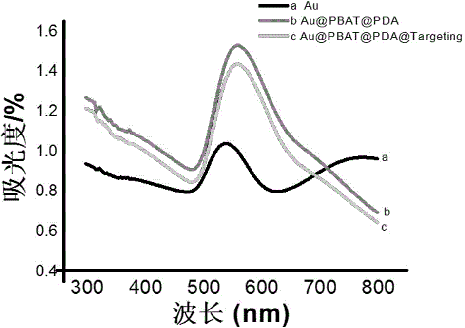
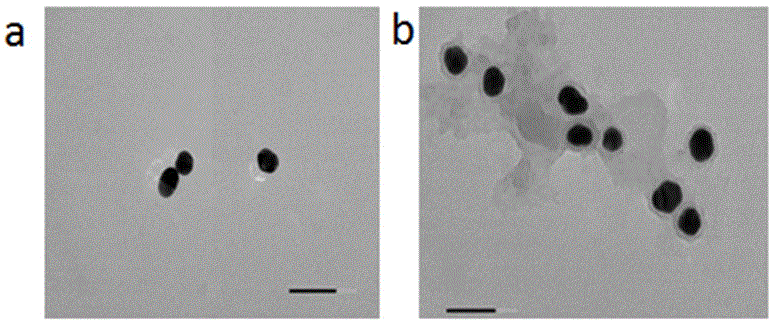
[0022] Example 1: Synthesis of surface-enhanced Raman scattering of human epidermal growth factor and gold-derived quinone-linked polypeptide for nuclear dopamine for imaging substrates of cell nuclei.

[0023]Pipette 1-2 mL of 10 mg / mL perchlorauric acid solution and 95-98 mL of deionized water into a 250 mL three-neck flask, mix them evenly, put them into an oil bath and heat, and control the Reflux once, add 750 uL of 1% trisodium citrate to it, keep reflux for about 20-30min, cool to room temperature, store it in a clean glass bottle, and store it in a refrigerator at 4°C. Take the synthesized 50-60 nm gold nanoparticles, centrifuge them at 8000-10000 rpm for 5-10 min, absorb the supernatant, redissolve it in the buffer solution, and add 5-10 uL of (E) -2-((4-(phenylethynyl)benzylidene)amino)ethanethiol, mix well and put it in the enzymolysis instrument to maintain the reaction for 1-2 min. Use the method of quinone group covalent coupling to modify the nuclear marker on ...

Embodiment 2

[0024] Example 2: Polypeptide-modified gold-nuclear Raman substrate imaging of the nucleus of glioma cell U251.

[0025] Add about 10 4 -10 5 Cells with a cell density of 18-24 hours are placed in a cell culture incubator, and 10-30 μg / mL of material is added to a glass dish, and the glass culture dish is placed in an incubator to interact with the cells for 18-24 hours. Aspirate the medium again, wash with PBS for 2-3 times, react with 4% paraformaldehyde solution in a 37°C incubator for 15-30 min, and finally wash with PBS for two to three times, and add 100-200 uL of PBS was stored in a refrigerator at 4°C and used for imaging of nuclei.


PUM


Abstract

The invention belongs to the technical field of biomacromolecular separation analysis and specifically relates to a surface enhanced raman scattering substrate for a gold nucleus 'cell raman silent zone', a preparation method and an application thereof in cell raman imaging. The method comprises the following steps: firstly, compounding 50-60nm gold nanoparticles according to a hydrothermal method and dissolving in a buffer solution; adding 'raman silent zone' molecule (E)-2-((4-(phenyl ethynyl) benzal) amino) ethanethiol and slightly agglomerating in the solution; adding dopamine into the buffer solution and connecting a polypeptide sequence with the golden ball surface through a quinonyl group generated by the auto polymerization of dopamine, thereby obtaining the surface enhanced raman scattering substrate for cell nucleus imaging of tumor cells. The material applied to the cell raman imaging has the advantages of high speed, high efficiency, simple operation and huge clinical application potentiality.

Description

technical field [0001] The invention belongs to the technical field of separation and analysis of biomacromolecules, and specifically relates to a surface-enhanced Raman scattering substrate with a gold-nucleated cell "Raman silent zone" and its preparation method and application in cell Raman imaging. Background technique [0002] Surface-enhanced Raman scattering is a special Raman spectral phenomenon, which is manifested on the rough surface of some gold, silver, copper and other precious metals. Geometric multiple (10 3 -10 9 )enlarge. This phenomenon greatly increases the sensitivity of Raman spectroscopy. At the same time, its smaller slit width and better spectral shape further expand its research in various fields. Since Raman's sensitivity is widely used in high-sensitivity analysis researches such as food safety, environmental monitoring, biology, medicine and archaeology, further exploration of its sensitivity has attracted attention from various directions. I...

Claims


Application Information

Patent Timeline
Patent Type & Authority Applications(China)
IPC IPC(8): G01N21/65B22F9/24
CPCG01N21/658B22F9/24
Inventor 高明霞陈萌张祥民
Owner FUDAN UNIV
Learn More