Fluorescence control method of light-controlled fluorescent protein marker biological tissue embedding sample

A fluorescent protein and control method technology, which is applied in the field of fluorescence control of light-controlled fluorescent protein-labeled biological tissue-embedded samples, can solve the problems of slow imaging of biological tissue-embedded samples, poor tomographic ability, and low axial resolution. Achieve the effect of convenient activation, precise control and good control effect

Active Publication Date: 2017-03-22
HUAZHONG UNIV OF SCI & TECH
View PDF6 Cites 2 Cited by
  • Summary
  • Abstract
  • Description
  • Claims
  • Application Information

AI Technical Summary

Problems solved by technology

[0003] Aiming at the above defects or improvement needs of the prior art, the present invention provides a fluorescence control method for labeling biological tissue-embedded samples with light-controlled fluorescent proteins, the purpose of which is to label biological tissue-embedded samples with light-sensitive fluorescent proteins Use specific wavelengths of activating light to activate the photoluminescent protein on the surface of the sample, and then excite the fluorescent protein on the surface of the sample with the corresponding wavelength of excitation light to realize the control of the surface fluorescence of the photoluminescent protein-labeled biological tissue-embedded sample, and by optimizing the fluorescence control condition to make its surface activation thickness thinner, and apply it to fluorescence tomography. Due to the lack of effective fluorescence control methods, due to the poor tomographic ability and low axial resolution during fast wide-field fluorescence imaging, the high-resolution point scanning imaging method is too slow for large-volume biological tissue-embedded samples.

Method used


Image

  • Fluorescence control method of light-controlled fluorescent protein marker biological tissue embedding sample
  • Fluorescence control method of light-controlled fluorescent protein marker biological tissue embedding sample
  • Fluorescence control method of light-controlled fluorescent protein marker biological tissue embedding sample

Examples


Embodiment 1

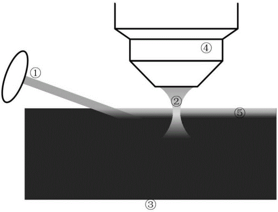
[0051] mEos3.1 is a common photoconvertible fluorescent protein. Its optical properties are that it only emits green fluorescence (excitation wavelength 488nm) and does not emit red fluorescence before being activated by ultraviolet 405nm wavelength; after being activated by ultraviolet 405nm wavelength for a short time, it emits red fluorescence under 561nm excitation light (emission spectrum is in above 550nm).

[0052] The method for embedding and light-sheet fluorescence control and fluorescence tomography of the mouse whole brain injected with Rv-dg-mEos3.1 in the cerebral cortex comprises the following steps:

[0053] (1) Sample fixation

[0054] The whole brain of the Rv-dg-mEos3.1-infected mouse was fixed by means of chemical fixation to obtain fixed mEos3.1-labeled biological tissue. Specific steps are as follows:

[0055] After the heart was perfused at 4°C, the dissected whole brain of the mouse was soaked in 4% PFA solution for about 12 hours. Use 40ml of PBS s...

Embodiment 2

[0069] The method for controlling fluorescence after embedding of Hela cells overexpressing PAGFP comprises the following steps:

[0070] (1) Sample fixation

[0071] Hela cells overexpressing PAGFP were fixed by chemical fixation means to obtain fixed PAGFP-labeled biological tissue. Specific steps are as follows:

[0072] At 4 degrees Celsius, Hela cells were soaked in 4% PFA solution for about 12 hours, and then rinsed with PBS solution for 3 times, using 40ml of PBS solution for each cell, and rinsed for 1 hour each time.

[0073] (2) Sample dehydration

[0074] The fixed PAGFP-labeled Hela cells were replaced with ethanol to dehydrate the sample to obtain a dehydrated PAGFP-labeled cell sample. The specific steps are:

[0075] At 4 degrees Celsius, the fixed PAGFP-labeled Hela cells were sequentially passed through 20 ml of gradient ethanol double-distilled water solution, soaked for 2 hours, and dehydrated. The concentration gradient of ethanol double distilled wate...

Embodiment 3

[0084] The method for controlling fluorescence after embedding of Hela cells overexpressing PAGFP comprises the following steps:

[0085] (1) Sample fixation

[0086] Hela cells overexpressing PAGFP were fixed by chemical fixation means to obtain fixed PAGFP-labeled biological tissue. Specific steps are as follows:

[0087] At 4 degrees Celsius, Hela cells were soaked in 4% PFA solution for about 12 hours, and then rinsed with PBS solution for 3 times, using 40ml of PBS solution for each cell, and rinsed for 1 hour each time.

[0088] (2) Sample dehydration

[0089] The fixed PAGFP-labeled Hela cells were replaced with ethanol to dehydrate the sample to obtain a dehydrated PAGFP-labeled cell sample. The specific steps are:

[0090] At 4 degrees Celsius, the fixed PAGFP-labeled Hela cells were sequentially passed through 20 ml of gradient ethanol double-distilled water solution, soaked for 2 hours, and dehydrated. The concentration gradient of ethanol double distilled wate...


PUM

PropertyMeasurementUnit
thicknessaaaaaaaaaa
angleaaaaaaaaaa
angleaaaaaaaaaa

Abstract

The invention discloses a fluorescence control method of a light-controlled fluorescent protein marker biological tissue embedding sample. Activation light is used for activating the light-controlled fluorescent protein on the surface layer of the sample, and exciting light is used for exciting the light-controlled fluorescent protein on the surface layer to emit fluorescence. The method comprises the following steps: selecting appropriate wavelength ranges of the activation light and the exciting light, and controlling an appropriate included angle between the activation direction and the upper surface of the sample at the same time, thereby realizing the precise control of the surface layer fluorescence of the light-controlled fluorescent protein marker biological tissue embedding sample, wherein the activation is convenient, and the thickness of the activated surface layer is thin. Only the fluorescence on the surface layer of the biological tissue embedding sample is activated when the fluorescence control method disclosed by the invention is used for activating; the exciting light excites the activated surface layer to perform fluorescence imaging when the method is applied to the fluorescent tomography of the biological tissue embedding sample, the thickness of the activated surface layer of the sample is the axial resolution of the fluorescence imaging, and the axial resolution is high.

Description

technical field [0001] The invention belongs to the field of fluorescence microscopic imaging, and in particular relates to a fluorescence control method for embedding samples of biological tissues marked with light-controlled fluorescent proteins. Background technique [0002] The photophysical properties of some fluorescent proteins can be precisely controlled by light, and these fluorescent proteins are called light-controlled fluorescent proteins. It includes at least the following two categories, photoactivatable fluorescent proteins and photoconversion fluorescent proteins. Light-activated fluorescent proteins do not fluoresce in the initial state, and can be excited to fluoresce after being activated by light at a specific wavelength, such as PAGFP. Photoconvertible fluorescent proteins can fluoresce in the initial state, but after being activated by light, they can be excited to fluoresce in another color, such as mEos3.1, mEos3.2, etc. The biological tissue labele...

Claims


Application Information

Patent Timeline
Patent Type & Authority Applications(China)
IPC IPC(8): G01N21/64
CPCG01N21/6486
Inventor 吕晓华刘秀丽张其郭文炎杨雄曾绍群
Owner HUAZHONG UNIV OF SCI & TECH
Why Eureka
Try Eureka