
A method of preparing a composite structure

A composite structure and construct technology, applied in the field of biology, can solve the problems of different culture systems, insufficient function, and disordered structure of artificial implants.

Active Publication Date: 2020-05-12
SICHUAN REVOTEK CO LTD
View PDF9 Cites 0 Cited by
  • Summary
  • Abstract
  • Description
  • Claims
  • Application Information

AI Technical Summary

Problems solved by technology

[0005] Although related technologies have been developed for many years, there are still significant deficiencies: 1. The culture process of MSCs induced differentiation is very cumbersome, and when MSCs are induced to differentiate into different types of cells, different culture systems are required
The entire cultivation process takes a long time, which greatly increases the risk of contamination
3. Using the method of planting seed cells on the surface of scaffold materials to construct artificial implants, it is difficult to accurately arrange the cells growing on the surface of the material, resulting in the formation of artificial implants with chaotic structures and insufficient functions

Method used


Image

  • A method of preparing a composite structure
  • A method of preparing a composite structure
  • A method of preparing a composite structure

Examples


specific Embodiment approach

[0285]The invention will now be described with reference to the following examples, which are intended to illustrate the invention, but not to limit it.

[0286] Reagents, kits or instruments whose sources are not specified in the examples are all conventional products commercially available on the market. Those skilled in the art understand that the examples describe the present invention by way of example and are not intended to limit the scope of the claimed invention.

Embodiment 1

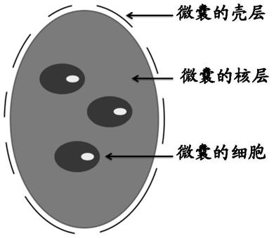
[0287] Embodiment 1. Preparation of microcapsules

[0288] This example provides an exemplary method for preparing microcapsules. The preparation method of microcapsules should be carried out under sterile conditions. In addition, if the microcapsules are intended to be applied to the human body, the biosafety level of the preparation method should reach the GMP workshop level.

[0289] The instrument used in this method is a granulator (BUCHI, Encapsulator, B-395 Pro), and the diameters of the concentric nozzles it is equipped with are as follows: the inner layer nozzle, 200 μm; the outer layer nozzle, 300 μm.

[0290] The materials used in this method are as follows:

[0291] (1) Materials used to prepare the nuclear layer

[0292] Type I collagen: 4mg / ml, neutralized with sterile 1M NaOH;

[0293] Sodium alginate: Dissolve and dilute with deionized water;

[0294] Vascular endothelial growth factor VEGF;

[0295] Type I collagen was mixed with 2% (w / v) (ie, 2 g / 100 ml...

Embodiment 2

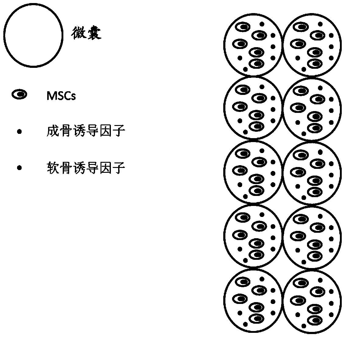
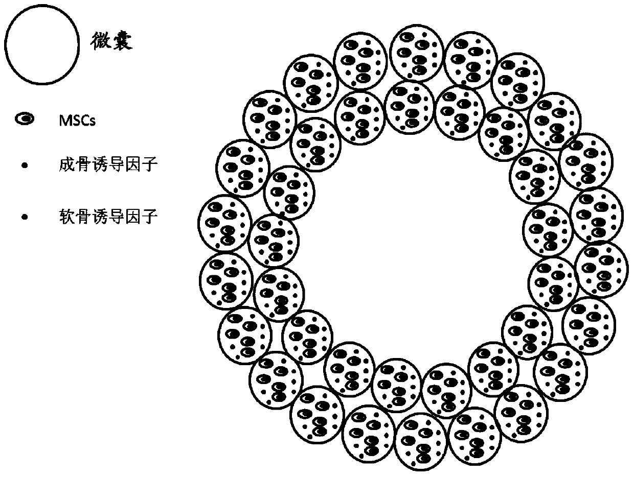
[0305] Example 2. Characterization of microcapsules

[0306] This example specifically analyzes the properties of the microcapsules prepared by the method in Example 1, including the size of the microcapsules, the thickness of the shell layer, the mechanical protection, the number of cells contained therein, and the like.

[0307] Use a microscope to observe the microcapsules prepared by the method of Example 1, the results are shown in Figures 3A-3C middle. Figures 3A-3C Micrographs of microcapsules prepared using a granulator under different instrument parameters (diameters of the inner and outer nozzles of the concentric nozzles) are shown, where, Figure 3A The diameter of the microcapsules in is about 120 μm (the scale bar is 100 μm); Figure 3B The diameter of the microcapsules in is about 200 μm (the scale bar is 100 μm); Figure 3C The diameter of the microcapsules in is about 450 μm (bar is 200 μm). These results suggest that the size of the microcapsules can be...


PUM

PropertyMeasurementUnit
elastic modulusaaaaaaaaaa
thicknessaaaaaaaaaa
elastic modulusaaaaaaaaaa

Abstract

Relating to the biology, regenerative medicine, bioprinting (like 3D bioprinting), tissue engineering and other technical fields, the invention particularly provides a method for preparation of a composite structure containing at least two cells, and the composite structure obtained by the method. In particular, preferably the composite structure provided by the invention is a composite structure comprising bones and cartilages, or a composite structure comprising endothelial cells and smooth muscle cells. Therefore, the invention provides a method for preparation of the composite structure containing osteocytes and chondrocytes, a method for preparation of the composite structure containing endothelial cells and smooth muscle cells, and a composite structure obtained by the method. In addition, the invention also relates to various applications of the composite structure.

Description

technical field [0001] The invention relates to the technical fields of biology, regenerative medicine, bioprinting (such as 3D bioprinting), tissue engineering and the like. In particular, the present invention provides a method for preparing a composite structure comprising at least two types of cells, and composite structures obtained by this method. Particularly preferably, the composite structure according to the invention is a composite structure comprising bone and cartilage, or a composite structure comprising endothelial cells and smooth muscle cells. Accordingly, the present invention provides a method of producing a composite structure comprising bone cells and chondrocytes, and a method of producing a composite structure comprising endothelial cells and smooth muscle cells, and composite structures obtained by said methods. Furthermore, the invention relates to various applications of the composite structures obtained. Background technique [0002] The vital st...

Claims


Application Information

Patent Timeline
Patent Type & Authority Patents(China)
IPC IPC(8): A61L27/38A61L27/40A61L27/58A61L27/54A61L27/50A61L27/24A61L27/20A61L27/18C12Q1/02G01N33/50
CPCA61L27/18A61L27/20A61L27/24A61L27/3808A61L27/3817A61L27/3821A61L27/3826A61L27/3834A61L27/50A61L27/54A61L27/58A61L2300/412A61L2430/02A61L2430/06G01N33/5008C08L5/04C08L3/02C08L75/04C08L77/04
Inventor 康裕建左潇
Owner SICHUAN REVOTEK CO LTD
Learn More