Method for screening and confirming kidney-yang deficiency animal model biomarker
A technology of biomarkers and animal models, applied in the field of screening and determination of biomarkers in animal models of kidney-yang deficiency syndrome, can solve the problems of lack of specificity and objectivity of evaluation indicators, and achieve the effect of promoting scientific evaluation and easy carrier.
- Summary
- Abstract
- Description
- Claims
- Application Information
AI Technical Summary
Problems solved by technology
Method used
Image
Examples
Embodiment 1
[0038] Establishment and identification of model rats with kidney-yang deficiency syndrome:
[0039] 1 Experimental consumables
[0040] 1.1 Experimental animals
[0041] A total of 40 male 3-month-old healthy SPF (Specific Pathogen Free) grade WISTAR rats, weighing 200-240 g, were purchased from Shanghai Slack Experimental Animal Co., Ltd. by the Experimental Animal Center of Fujian University of Traditional Chinese Medicine. The experimental animal license number: SCXK (Shanghai) 2012-0002, laboratory animal quality certificate number: 2007000548015. Experimental animal use license number of the Experimental Animal Center of Fujian University of Traditional Chinese Medicine: SYXK (Fujian) 2014-0001.
[0042] 1.2 Main reagents and drugs
[0043] Table 1 Main reagents and drugs
[0044]
[0045] 1.3 Main Instruments
[0046] Table 2 Main Instruments
[0047]
[0048] 2 Experimental methods
[0049] 2.1 Animal grouping
[0050] Forty WISTAR rats were adaptively fe...
Embodiment 2
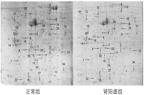
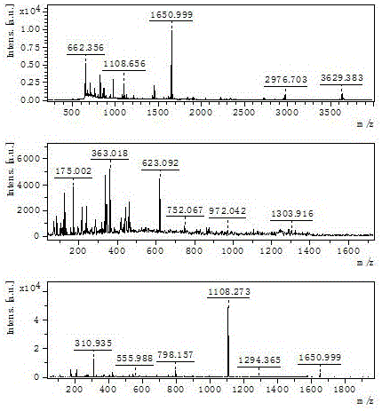
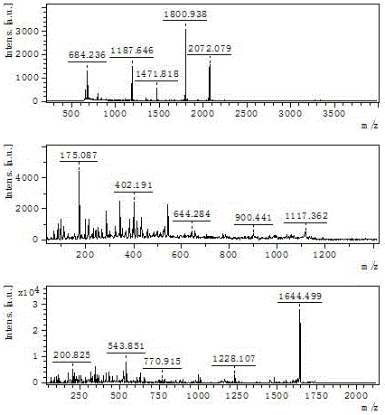
[0063] Screening of differential proteins, which includes the following steps:
[0064] 1. Extraction of cancellous bone total protein
[0065] ①Cut the frozen fresh femur stem with bone scissors, rinse the bone marrow with ultrapure water, and gently scrape the dry cancellous bone of the femur into the mortar (pre-cooled with liquid nitrogen) with a curette, and add Liquid nitrogen and rapid grinding, repeated grinding 3 to 5 times, the powder was taken and weighed, then protein sample lysate was added according to the ratio of 1 g bone tissue to 1.8 mL lysate, and the protein was lysed and extracted in a refrigerator at 4°C for 4 h (Mix thoroughly every 1 hour); ②Place the EP tube containing the bone tissue suspension in a low-temperature high-speed centrifuge, centrifuge at 100,000 rpm at 4°C for 1 hour, and absorb the supernatant; ③Add nuclease: Add 2 ulDNase and 2 ul RNase to 1 ml of the lysate, and place on ice for 15 min; ④ Ultrasound: use an ultrasonic cleaner to soni...
Embodiment 3
[0092] Western Blot and RT-PCR technology double verification from protein and gene level, it includes the following steps:
[0093] 1. Western Blot detection of cancellous bone-related protein in the femoral shaft of rats in each group
[0094] 1.1 Extraction of cancellous bone total protein
[0095] The cancellous femur bone of 8 rats in each group was randomly selected from the -80°C freezer, placed on ice, and the bone tissue was quickly cut into pieces with a rongeur, put into a pre-cooled grinder, and quickly ground to In powder form, liquid nitrogen was added to cool at the right time during the grinding process, then transferred to an Eppendorf tube, and weighed; 1 g of bone tissue was added to 1 ml of lysate according to the proportion, oscillated by a micro-vortexer for 5 s, and then placed in a refrigerator at 4 °C. Vortex once every 30 min, take it out after 4 h; transfer to a low-temperature high-speed centrifuge, set the program at 4 °C, 14000 rpm, centrifuge fo...
PUM
 Abstract
Description
Claims
Application Information
 - R&D
- Intellectual Property
- Life Sciences
- Materials
- Tech Scout
- Unparalleled Data Quality
- Higher Quality Content
- 60% Fewer Hallucinations
Browse by: Latest US Patents, China's latest patents, Technical Efficacy Thesaurus, Application Domain, Technology Topic, Popular Technical Reports.
© 2025 PatSnap. All rights reserved.Legal|Privacy policy|Modern Slavery Act Transparency Statement|Sitemap|About US| Contact US: help@patsnap.com


