
Method for preparing internal hemostatic dressing by programmed cooling

A technology of hemostatic dressing and cooling rate, which is applied in the field of preparation of hemostatic dressings in vivo, can solve the problems of affecting the hemostatic material's absorption of oozing blood on the wound surface, long clinical hemostasis time of sponge, uneven surface of hemostatic material, etc. Hidden danger, difficult secondary bleeding, and the effect of improving the hemostatic effect

Active Publication Date: 2020-06-09
NORTHWEST UNIV
View PDF2 Cites 0 Cited by
  • Summary
  • Abstract
  • Description
  • Claims
  • Application Information

AI Technical Summary

Problems solved by technology

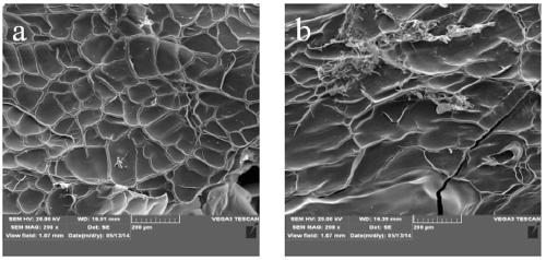
[0003] At present, vacuum freeze-drying is the most widely used method in the preparation of collagen hemostatic materials. However, the process of pre-freezing before vacuum freeze-drying has not been deeply studied and optimized, resulting in uneven surface of the prepared hemostatic material. The defects in physical and chemical properties, such as uneven pore size and obvious difference between the upper and lower surfaces, affect the absorption of hemostatic material on wound oozing blood, as well as the interaction with platelets and coagulation factors in the blood, resulting in a longer clinical hemostasis time and poor efficacy of the final sponge , such as patent CN 200610041913

Method used


Image

  • Method for preparing internal hemostatic dressing by programmed cooling
  • Method for preparing internal hemostatic dressing by programmed cooling
  • Method for preparing internal hemostatic dressing by programmed cooling

Examples


preparation example Construction

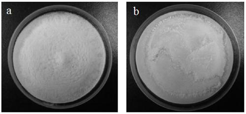
[0027] A preparation method of an internal hemostatic dressing, comprising the following steps:

[0028] Step 1: Dissolve humanoid collagen in water for injection, stir to dissolve it completely, so that the mass concentration of humanoid collagen in the solution is 1%-5%. A human-derived collagen produced by high-density fermentation;

[0029] Step 2: Transfer the solution into a disposable sterile Petri dish or mold, 7-15 mL per dish or mold;

[0030] Step 3: Use the program freezer to control the cooling rate at 4-8°C / min during the pre-freezing process for precise cooling down to -40--80°C;

[0031] Step 4: Freeze-dry the sample in a vacuum freeze dryer for 24-48 h;

[0032] Step 5: Place the freeze-dried sample in a vacuum drying oven for cross-linking, adjust the cross-linking temperature to 100°C-150°C, and the cross-linking time to 24h-96h;

[0033] Step 6: Sterilize by irradiation for 10-30 hours.

Embodiment 1

[0035] Dissolve the human-like collagen in water for injection, stir to dissolve it completely, so that the mass concentration of the human-like collagen in the mixed solution is 1%, and then place it in a programmed freezer to freeze at a rate of -4°C / min to -80°C and kept for 2 h, quickly transferred to a vacuum freeze dryer to freeze-dry for 24 h, then transferred to a vacuum oven at 100°C for cross-linking for 24 h, took out the package and irradiated with Co60 and sterilized for 24 h to obtain Internal hemostatic dressing.

Embodiment 2

[0037] Dissolve human-like collagen in water for injection, stir to dissolve completely, so that the mass concentration of human-like collagen in the mixed solution is 2%, and then place it in a programmed freezer at a rate of -4°C / min to - 80°C and kept for 2 h, quickly transferred to a vacuum freeze dryer to freeze-dry for 40 h, then transferred to a vacuum drying oven at 110°C for cross-linking for 28 h, took out the package for Co60 irradiation cross-linking and sterilized for 24 h , to obtain internal hemostatic dressings.


PUM


Abstract

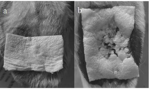
This patent discloses a method for preparing a hemostatic dressing in the body. The preparation method uses human-like collagen as raw material, adds water for injection, stirs and dissolves, then freezes by setting a specific cooling rate to achieve precise cooling and pre-freezing, and vacuum freeze-drying. , and then after thermal cross-linking, Co60 irradiation sterilization, and finally an in-vivo hemostatic dressing was obtained. Preparation method of the present invention The in-vivo hemostatic dressing prepared by the present invention has high safety, good biocompatibility, and significant hemostatic effect. It can be used to stop bleeding on various wounds and after surgery, and has good application in related fields such as medical biomaterials and tissue engineering. application prospects.

Description

technical field [0001] The invention relates to a method for preparing a hemostatic dressing in the body, in particular to a method for preparing the hemostatic dressing in the body through programmed cooling, and belongs to the field of biomedical materials. Background technique [0002] Collagen is a high-molecular protein that is generally white, opaque, and fibrous. It is one of the main components of the structure of organisms and is widely distributed in the skin, bones and muscle tissues of animals. Collagen has hemostatic properties. On the one hand, it can strengthen the adhesion and aggregation of platelets to form thrombus and prevent blood from flowing out. On the other hand, the intrinsic coagulation pathway can also be initiated by activating and inducing various coagulation factors. However, collagen is mostly derived from animal skin, tendons, etc., and its processability is poor. During the extraction process, part of the biological activity of the protein...

Claims


Application Information

Patent Timeline
Patent Type & Authority Patents(China)
IPC IPC(8): A61L26/00A61L24/10A61L24/00
CPCA61L24/0036A61L24/102A61L26/0033A61L26/0085A61L2400/04C08L89/00
Inventor 范代娣米钰惠俊峰马晓轩马沛段志广刘琳
Owner NORTHWEST UNIV
Learn More