Method for measuring hydrolytic enzyme activity

A hydrolytic enzyme and active technology, applied in the field of biological analysis, can solve the problems of complex probe preparation process, insufficient anti-interference ability, poor anti-interference ability, etc., and achieve good reproducibility, short detection time, and strong anti-interference ability Effect

Inactive Publication Date: 2017-05-31
NANJING UNIV OF SCI & TECH
View PDF4 Cites 0 Cited by
  • Summary
  • Abstract
  • Description
  • Claims
  • Application Information

AI Technical Summary

Problems solved by technology

Taking β-galactosidase as an example, there are two types of colorimetric analysis methods currently available for its activity determination: (1) p-nitrophenyl-β-D-galactopyranoside (or o-nitro Phenyl-β-D-galactopyranoside) is the chromogenic substrate, because β-galactosidase can hydrolyze it to generate p-nitrophenol (or o-nitrophenol), the latter solution is in alkaline It is yellow under certain conditions and has a maximum absorption peak at 405nm (or 420nm), thereby realizing the colorimetric analysis of β-galactosidase activity, but there are defects such as poor selectivity, low sensitivity, narrow detection range and insufficient anti-interference ability ; Moreover, due to the poor stability of the substrate, it is easy to cause "false positive" results (1.Maruhn D.(1976).Rapid colorimetric assay ofβ-galactosidase and N-acetyl-β-glucosaminidase in human urine[J].ClinicaChimica Acta, 73(3):453-461; 2. Gary, R.K., Kindell, S.M.(2005). Quantitative assay of senescence-associated β-galactosidase activity in mammalian cellextracts. Analytical Biochemistry, 343(2), 329-334.); (2 ) uses gold nanoparticles as probes, based on the property that the color of the solution changes due to the change of the aggregation state of gold nanoparticles, and realizes the colorimetric analysis of the activity of β-galactosidase, but the preparation process of the probe is complicated, the detection period is long, and the cost High and poor anti-interference ability and other defects (Zeng Z., etal. (2012). Simple and real-time colorimetric assay for glycosidases activity using functionalized gold nanoparticles and its application for inhibitorscreening. Analytical Chemistry, 84 (21), 9089-9095; Chen J., et al. (2016). Colorimetric Detection of Escherichia coli Based on the Enzyme-Induced Metallization of Gold Nanorods. Small, 12(18), 2469-2475.)

Method used


Image

  • Method for measuring hydrolytic enzyme activity
  • Method for measuring hydrolytic enzyme activity
  • Method for measuring hydrolytic enzyme activity

Examples


Embodiment 1

[0033] The method of the invention is used in a feasibility analysis experiment for measuring the activity of β-galactosidase.

[0034] 10μL 0.01mg·mL -1 β-galactosidase solution was added to 590 μL containing 10 mM p-aminophenyl-β-D-galactopyranoside, 0.25 mM Fe 3+ and 0.75 mM bathophenanthroline sodium disulfonate in the detection solution, after incubation at 25° C. for 15 min, record the ultraviolet-visible absorption spectrum of the detection solution.

[0035] In feasibility analysis experiments, missing components were replaced with the same volume of buffer. From figure 1 It can be seen that when all components exist (a), the detection solution has a strong absorption peak at ~535nm; and when no β-galactosidase (b), p-aminobenzene Base-β-D-galactopyranoside (c), Fe 3+ (d), or sodium bathophenanthroline disulfonate (e), its absorption value at ~535nm is very low. It can be seen that the method of the present invention can be used to measure the activity of β-galact...

Embodiment 2

[0037] The method of the invention is used for determining the relationship between the detection signal of the β-galactosidase activity and the β-galactosidase activity.

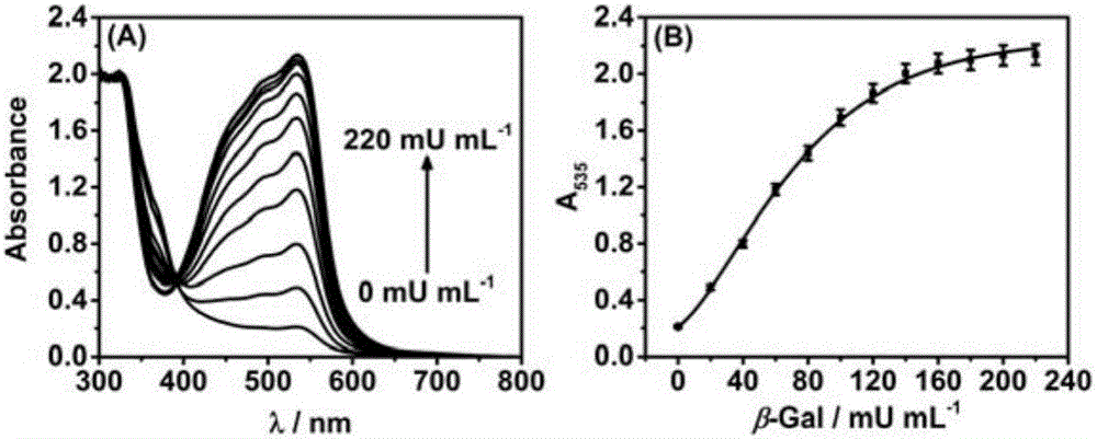
[0038] 10 μL of 0, 20, 40, 60, 80, 100, 120, 140, 160, 180, 200 and 220 mU·mL -1 The β-galactosidase solution was added to 590 μL containing 10 mM p-aminophenyl-β-D-galactopyranoside, 0.25 mM Fe 3+ and 0.75 mM bathophenanthroline sodium disulfonate in the detection solution, after incubation at 25° C. for 15 min, record the ultraviolet-visible absorption spectrum of the detection solution.

[0039] From figure 2 It can be seen that with the increase of β-galactosidase activity, the absorption value of the detection solution at ~535nm increases correspondingly, and the absorption intensity has a good quantitative relationship with β-galactosidase activity. It can be seen that the method of the present invention can be used for quantitative determination of β-galactosidase activity.

Embodiment 3

[0041] The method of the invention is used to determine the selectivity of β-galactosidase activity.

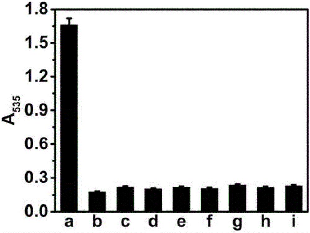
[0042] 10μL 0.01mg·mL -1 β-galactosidase or 10μL 0.1mg·mL -1 The other protein solution was added to 590 μL containing 10 mM p-aminophenyl-β-D-galactopyranoside, 0.25 mM Fe 3+ and 0.75 mM bathophenanthroline sodium disulfonate in the detection solution, after incubation at 25° C. for 15 min, record the absorption value of the detection solution at ~535 nm.

[0043] From image 3 It can be seen that only when β-galactosidase is added, the absorption value of the detection solution at ~535nm will increase significantly. It can be seen that the method of the present invention has high selectivity when used for measuring the activity of β-galactosidase.


PUM


Abstract

The invention discloses a method for measuring hydrolytic enzyme activity. The method comprises the following steps: incubating a detection solution containing a substrate, Fe<3+> and bathophenanthrolinedisulfonic acid disodium salt with a hydrolase solution to be measured; making the substrate react with hydrolase to generate a reduction product; reducing the Fe<3+> into Fe<2+> by the reduction product; combining the Fe<2+> and the bathophenanthrolinedisulfonic acid disodium salt to form a Fe<2+>-bathophenanthroline compound, wherein the detection solution turns from colorless to crimson, and has a very strong absorption peak at 535nm. The higher the hydrolytic enzyme activity in a sample is, the deeper the color of the detection solution becomes. The method has high detection sensitivity for the hydrolytic enzyme activity, high selectivity, high interference resistance, good reproducibility and short detection time, is easy and convenient to operate, and can be applied to rapid and high-sensitivity detection of the hydrolytic enzyme activity in a clinical sample; the detection cost is lowered greatly.

Description

technical field [0001] The invention belongs to the technical field of biological analysis, and relates to a method for measuring hydrolase activity, in particular to a method for measuring hydrolase activity based on colorimetric analysis. Background technique [0002] Hydrolase is a general term for a class of enzymes that catalyze hydrolysis reactions, and widely exists in various animals, plants and microorganisms. Simple, rapid and sensitive determination of hydrolase activity is of great significance for early diagnosis of diseases, observation of curative effect and monitoring of prognosis. [0003] At present, fluorescence analysis and colorimetric analysis are mostly used for the determination of hydrolytic enzyme activity. Among them, colorimetric analysis, also known as absorptiometry, is based on the change of the color of the solution, that is, the selective absorption of light by the solution, to quantitatively analyze the content of the substance. It has the ...

Claims


Application Information

Patent Timeline
Patent Type & Authority Applications(China)
IPC IPC(8): G01N21/31
CPCG01N21/314G01N2021/3155
Inventor 孔金明胡琼何玟辉梅亚琦张学记
Owner NANJING UNIV OF SCI & TECH
Why Eureka
Try Eureka