Crystalline lens biomechanics and optical property noninvasive in-vivo imaging system and measuring method

A technology of optical characteristics and biomechanics, applied in medical science, equipment for testing eyes, gonioscopy, etc., can solve problems such as limited imaging depth, insufficient information, and limited research and development, and achieve accurate and safe results High, easy-to-operate effect

CN106963338AActive Publication Date: 2017-07-21浙江瑞瞳生物科技有限公司
8 Cites 20 Cited by

Patent Information

Authority / Receiving Office
CN · China
Current Assignee / Owner
Publication Date
2017-07-21

Smart Images

  • Figure 1
    Figure 1
  • Figure 2
    Figure 2
  • Figure 3
    Figure 3
Patent Text Reader

Abstract

The invention discloses a crystalline lens biomechanics and optical property noninvasive in-vivo imaging system and a measuring method. The ultrasonic load system provides mechanical waves capable of enabling a crystalline lens to deform, a spectral domain OCT system achieves the structure and elastic imaging of the crystalline lens; on the basis of the yoke eliminating technology, OCT full-range imaging is achieved, full anterior segment two-dimensional or three-dimensional structure images from the anterior corneal surface to the retrolental surface, morphological and optical characteristic parameters of the crystalline lens are obtained through calculation, and a crystalline lens topographic map is constructed. Meanwhile, the phase sensitive OCT technology is utilized, strain distribution in the depth direction of the crystalline lens is obtained, and a two-dimensional or three-dimensional biomechanical characteristic distribution diagram of the crystalline lens is obtained through rebuilding. High-resolution imaging and precision measurement of biomechanics and optical properties of the crystalline lens provide the more comprehensive and complete imaging surveying basis for fundamental research, early diagnosis, operation plan formulating and postoperation result evaluation of cataract and other crystalline lens diseases, and the important practical significance and clinical significance are achieved.
Need to check novelty before this filing date? Find Prior Art

Description

technical field

[0001] The present invention relates to lens optical coherence elastography and full-range imaging optical coherence tomography technology, in particular to a method of using spectral domain optical coherence tomography technology combined with phase-sensitive detection technology to analyze the biomechanical and optical properties of the lens Systems and methods for non-invasive in vivo imaging and measurement. Background technique

[0002] The lens is one of the important refractive media of the eye. Physiological and pathological changes in its biomechanical and optical properties (such as elasticity, refractive power, and refractive index, etc.) will lead to the occurrence and development of many ophthalmic diseases, such as presbyopia and cataract. Cataract is the world's first blinding eye disease. Aging is its most common etiology and the incidence increases with age. As the life expectancy of the population increases, the social impact of cataracts ...

Examples

Embodiment Construction

[0036] The present invention will be further described below in conjunction with the accompanying drawings and examples, but the protection scope of the present invention is not limited to the following examples.

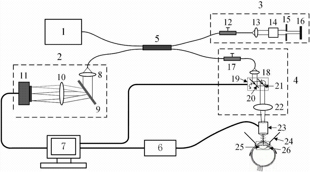
[0037] like figure 1 As shown, the noninvasive in vivo imaging system for lens biomechanics and optical properties includes a light source 1, a spectrometer 2, a reference arm 3, a sample arm 4, an ultrasonic loading system and a control computer 7; the light source 1 uses a near-infrared broadband light source. The optical signal emitted by the light source 1 is divided into two paths through the fiber coupler 5 : one path enters the sample arm 4 , and the other path enters the reference arm 3 . The ultrasonic loading system consists of an ultrasonic probe 23 and a pulse generator 6 .

[0038] The sample arm 4 is composed of a first polarization controller 17 , a first collimator lens 18 , an X-Y scanning galvanometer system 19 and a scanning lens 22 . In the sam...