Focusing photoinduced ultrasound material, preparation method thereof and endoscopic photoinduced ultrasound probe

A technology of photoinduced ultrasound and focused light, which is applied in ultrasound therapy, ultrasound/sonic/infrasonic diagnosis, diagnostic probe accessories, etc., and can solve the problems of large volume and low accuracy of piezoelectric ultrasonic transducers

Inactive Publication Date: 2017-08-18
HUAZHONG UNIV OF SCI & TECH
View PDF10 Cites 3 Cited by
  • Summary
  • Abstract
  • Description
  • Claims
  • Application Information

AI Technical Summary

Problems solved by technology

[0006] In view of the above defects or improvement needs of the prior art, the present invention provides a focused photo-induced ultrasonic material and its manufacturing method and an endoscopic photo-induced ultrasonic probe, the purpose of which is to make the probe smaller in size, higher in accuracy, and Diagnose and treat at the same time, thereby solving the technical problems of large volume and low accuracy of piezoelectric ultrasonic transducers

Method used


Image

  • Focusing photoinduced ultrasound material, preparation method thereof and endoscopic photoinduced ultrasound probe
  • Focusing photoinduced ultrasound material, preparation method thereof and endoscopic photoinduced ultrasound probe
  • Focusing photoinduced ultrasound material, preparation method thereof and endoscopic photoinduced ultrasound probe

Examples


Embodiment 1

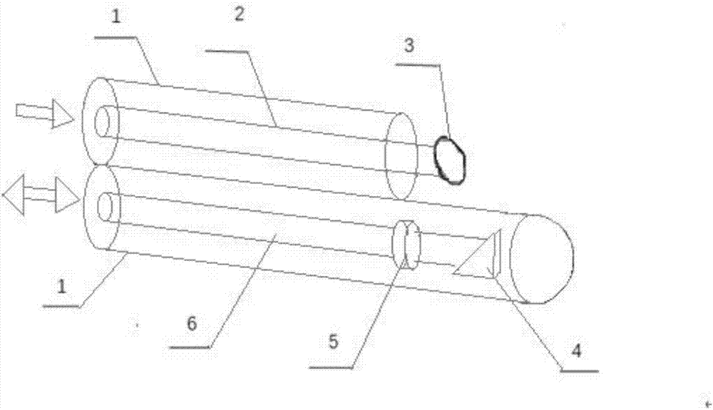
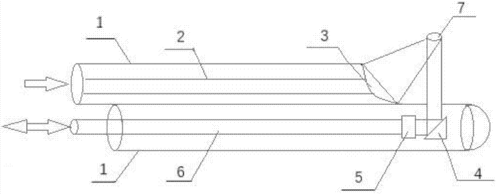
[0042] figure 1 is a schematic diagram of the three-dimensional structure of the endoscopic photoinduced ultrasound probe, figure 2 is a cross-sectional view of an endoscopic photo-ultrasound probe. First, the imaging incident fiber (6) is bonded together with the cylindrical photoinduced ultrasonic material (5). The diameter of the cylindrical photoinduced ultrasonic material (5) is 2 mm. The center of circle of material (5) is on the same straight line. Then, the therapeutic incident optical fiber (2) and the focused photo-induced ultrasonic material (3) are bonded together, the focused photo-induced ultrasonic material (3) has a diameter of 2 millimeters, and there is no gap between the two. The total reflection mirror (4) is fixed on the right side of the cylindrical photo-ultrasonic material (5), and the axis of the total reflection mirror (4) is on the same line as the center of the cylindrical photo-ultrasonic material (5). A layer of casing (1) is respectively wrap...

Embodiment 2

[0045] figure 1 is the schematic diagram of the three-dimensional structure of the photoinduced ultrasound probe, figure 2 is a cross-sectional view of the photo-ultrasonic probe. First, the imaging incident fiber (6) is bonded together with the cylindrical photoinduced ultrasonic material (5). The diameter of the cylindrical photoinduced ultrasonic material (5) is 3 mm. The center of circle of material (5) is on the same straight line. Then, the therapeutic incident optical fiber (2) and the focused photo-induced ultrasonic material (3) are bonded together. The focused photo-induced ultrasonic material (3) has a diameter of 5 mm, and there is no gap between the two. The total reflection mirror (4) is fixed on the right side of the cylindrical photo-ultrasonic material (5), and the axis of the total reflection mirror (4) is on the same line as the center of the cylindrical photo-ultrasonic material (5). A layer of casing (1) is respectively wrapped outside the two incident...

Embodiment 3

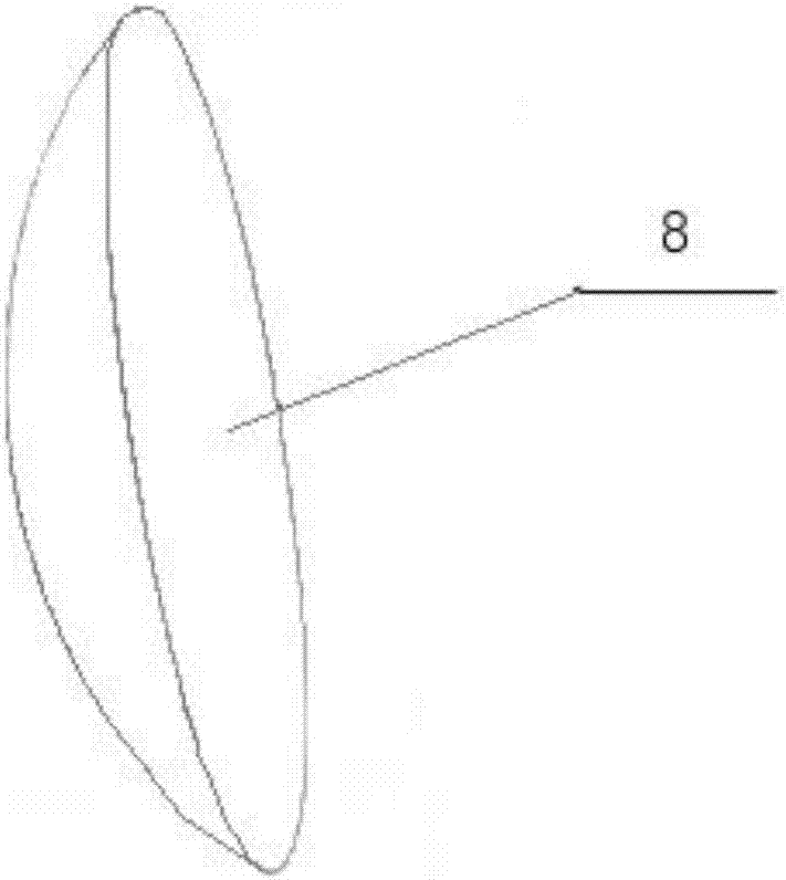
[0048] image 3 is a schematic diagram of the three-dimensional structure of the focused photo-induced ultrasound material (3), Figure 4 is a schematic diagram of the production of the focused photo-ultrasonic material (3), Figure 5 is a schematic diagram of the structure of a stereolithography apparatus based on mask image projection. The fabrication of the focused photo-ultrasonic material (3) is divided into two steps. The first step is to prepare the azeotropic mixture of methyl ethyl ketone and ethanol, and use the stainless steel grinding balls in the planetary ball mill to grind the carbon nanotube powder and the dispersant polyvinyl alcohol in the azeotropic mixture through the planetary ball mill for 10 hours. The speed is 150rpm. The mixture was then dried at 40°C for 11 hours. After evaporation of the solvent in the dispersion, a dry carbon nanotube powder can be obtained. In the second step, the carbon nanotube powder obtained in the first step is mixed with...


PUM

PropertyMeasurementUnit
diameteraaaaaaaaaa
diameteraaaaaaaaaa
diameteraaaaaaaaaa

Abstract

The invention provides a focusing photoinduced ultrasound material, a preparation method thereof and an endoscopic photoinduced ultrasound probe. The focusing photoinduced ultrasound material is prepared layer by layer from bottom to top taking a mixture of carbon nanotube powder and a light-cured resin as a raw material by utilizing the 3D printing technology based on mask image projection stereo lithography technology (MIPS). Main structure of the endoscopic photoinduced ultrasound probe comprises an imaging incident optical fiber, a treatment incident optical fiber, a cylindrical photoinduced ultrasound material, the focusing photoinduced ultrasound material and a completely reflecting mirror. Through the imaging incident optical fiber and the cylindrical photoinduced ultrasound material, an ultrasonic signal is generated, and a position of a lesion tissue is determined; and then, through the treatment incident optical fiber and the focusing photoinduced ultrasound material, an ultrasonic signal having high energy is generated, and the lesion tissue is smashed.

Description

technical field [0001] The invention belongs to the field of ultrasonic energy conversion, and more specifically relates to a focused photo-induced ultrasonic material, a preparation method thereof, and an endoscopic photo-induced ultrasonic probe. Background technique [0002] With the development of society and economy, the incidence of cardiovascular diseases in my country is showing an increasing trend year by year. Early diagnosis and treatment of diseased tissues is the key to overcoming cardiovascular diseases. Currently, diseased tissue can be diagnosed using existing technologies. There are researches and reports on the use of piezoelectric ultrasonic transducers for diagnosis and treatment of intravascular diseased tissue. High-frequency ultrasound is used for imaging and low-frequency ultrasound is used for treatment. With the development of piezoelectric single crystal and thick film technology, miniature high-frequency ultrasonic transducers can improve the qua...

Claims


Application Information

Patent Timeline
Patent Type & Authority Applications(China)
IPC IPC(8): A61N7/00B29C64/135B29C64/314B33Y40/00
CPCB33Y40/00A61N7/00A61N2007/0043A61N2007/0004A61N2007/0056A61N2007/0052A61B8/4444B33Y80/00A61N7/02A61N7/022A61B2017/00526A61N2007/0065A61B8/42B29C64/135B29C64/314
Inventor 朱本鹏申明柯杨晓非张悦欧阳君
Owner HUAZHONG UNIV OF SCI & TECH
Why Eureka
Try Eureka